- Center on Health Equity & Access
- Clinical
- Health Care Cost
- Health Care Delivery
- Insurance
- Policy
- Technology
- Value-Based Care
Radiomics on Spinal Cord MRI May Offer Superior MS Assessment
Radiomic assessment has been used to distinguish multiple sclerosis from similar disorders, and it might also be useful in identifying MS stages and subtypes.
Radiomics may offer a superior means to assess spinal cord pathology compared with visual assessment of MRI in patients with multiple sclerosis (MS), a new report found.
The findings may make it easier for clinicians to more precisely understand how the disease is progressing in patients. The report was published in the Journal of Neuroimaging.1
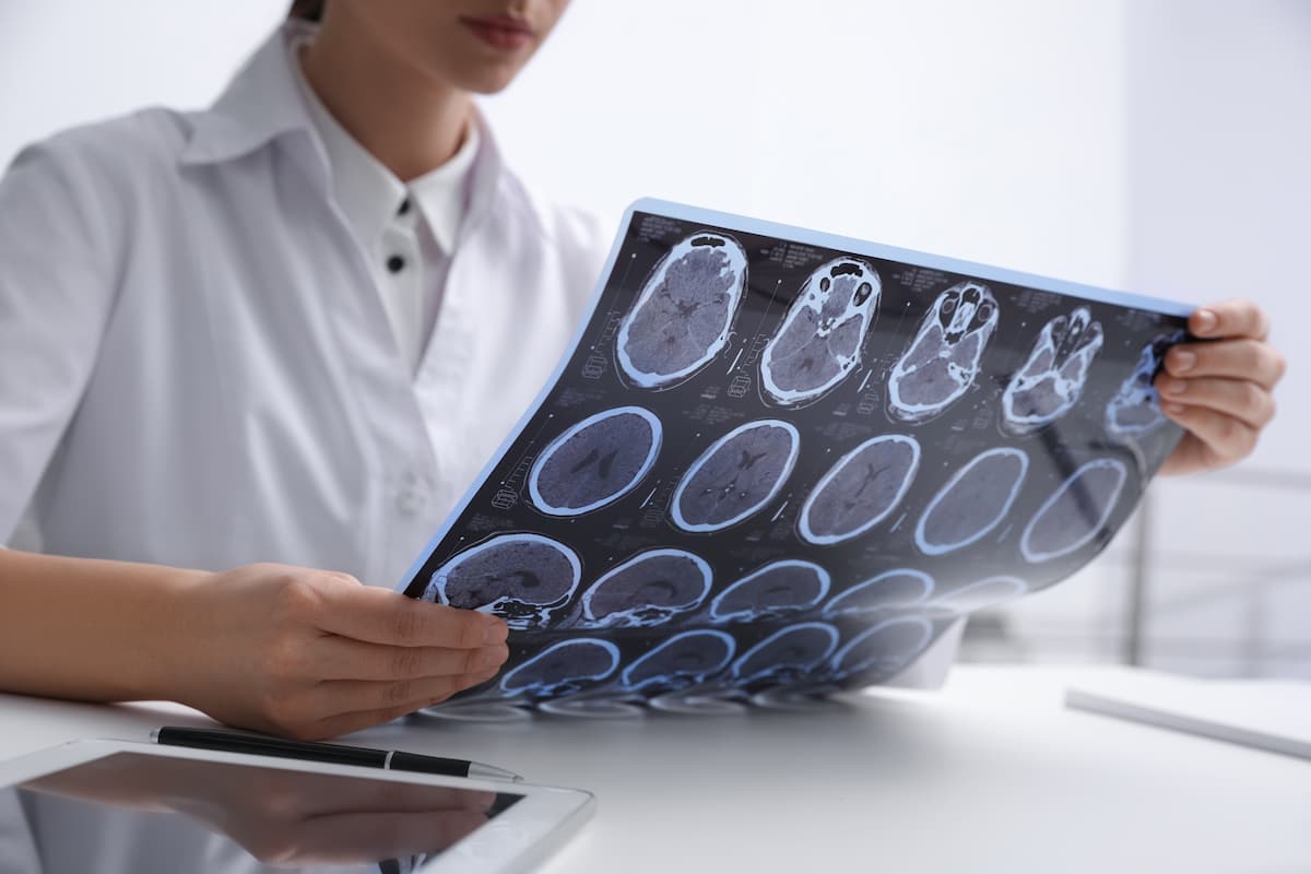
The investigators identified features that differentiate MS by relapsing vs progressive course, as well as by subgroups known to be associated with distinct pathologies, such as age and race. | Image credit: New Africa - stock.adobe.com

MRI has long played a role in the treatment of MS, the authors noted.
“Advances in brain MRI have facilitated visualization and recapitulation of neuroinflammatory pathology in people with multiple sclerosis (pwMS),” they wrote. “Despite this, there remains a large discrepancy between the degree of disease activity on MRI and clinical disability.”
That discrepancy, they said, suggests a limit to the utility of conventional MRI in explaining global disability.
One potential way to refine disability tracking is to look at spinal cord pathology, which the authors said is both common in MS and “highly relevant” to disability. The problem, they said, is that conventional spinal cord MRI “lacks sensitivity and reliability, largely related to technical issues given the small caliber of the cord and associated physiological mobility.”
A potential solution, they said, is radiomics, which uses signal intensities and interrelationships between pixels to extract features from images.
“The detected heterogeneity within regions of interest (ROI) is thought to reflect physiological and pathological changes in tissues, many of which may be undetectable by traditional visual inspection,” they said.
For instance, a 2022 study found that radiomic signatures could be used to differentiate lesions associated with MS from similar lesions related to ischemic demyelination, which the authors said often mimics MS.2
While there is significant evidence that radiomics can be used to distinguish between MS and other demyelinating disorders, there is relatively little evidence related to whether radiomics can be used to distinguish between subgroups of people with MS and track disease progression, the authors explained.1
The authors conducted a retrospective, cross-sectional cohort study of 2966 people with MS who were at least 16 years old and underwent MRI and clinical assessment within 3 months. All of the patients were treated at Cleveland Clinic centers between 2015 and 2022. The investigators also created a cohort of 41 age- and sex-matched controls who had undergone MRI studies using the same protocols as the patients with MS.
The investigators extracted radiomic features from upper cervical cord coverage on cross-sectional 3.0T brain non-contrasted T1-weighted MRI.
Altogether, 93 radiomic features were analyzed, and the authors found that 12 of those features differed significantly between patients with MS and healthy controls. Four features were of the gray-level co-occurrence matrix (GLCM) class, and the authors said those features were also associated with disability measures on assessments such as the Patient-Determined Disease Steps (PDDS) assessment, the manual dexterity test (MDT), and the walking speed test (WST).
“Each GLCM feature that was higher among pwMS with worse disability (i.e., higher PDDS and lower WST/MDT), and vice versa,” the authors said. PDDS was found to have the most correlation with radiomic features, and a smaller number of correlations were identified with WST and MDT, the authors said.
The researchers also identified features that differentiate MS by relapsing versus progressive course, as well as by subgroups known to be associated with distinct pathologies, such as age and race.
Radiomic spinal cord assessment outperformed assessment of conventional spinal cord MRI features in the study.
The authors said one challenge in utilizing radiomics is translating its mathematical output into clinically meaningful data. They said more studies should be undertaken to refine radiomics as it applies to MS.
“This may result in recognition of a more clearly defined and biologically based representation of radiomic features associated with clinical course and disability outcomes,” they wrote. In the meantime, they said their findings support the idea that radiomics might become an important tool in the evaluation and treatment of MS.
References
1. Lambe J, Thompson NR, Li Y, Nakamura K, Ontaneda D. Association of spinal cord radiomic features and disability in multiple sclerosis. J Neuroimaging. 2025;35(5):e70089. doi:10.1111/jon.70089
2. He T, Zhao W, Mao Y, et al. MS or not MS: T2-weighted imaging (T2WI)-based radiomic findings distinguish MS from its mimics. Mult Scler Relat Disord. 2022;61:103756. doi:10.1016/j.msard.2022.103756
