- Center on Health Equity & Access
- Clinical
- Health Care Cost
- Health Care Delivery
- Insurance
- Policy
- Technology
- Value-Based Care
Nanomechanical Signatures May Boost Pulmonary Fibrosis Diagnosis, Monitoring
Atomic force microscopy successfully identified nanomechanical changes in fibrotic lung tissue.
Atomic force microscopy (AFM) may be a valuable tool to help clinicians more closely stage pulmonary fibrosis (PF), according to a new report. The study, which was published in the journal Small, also suggests that the tool can be used to track response to therapy and personalize care.1
Andreas Stylianou, MSc, PhD, of the European University Cyprus, explained along with colleagues that one of the struggles in treating pulmonary fibrosis is that the current standard-of-care therapies have highly variable efficacy.
“This variability in effectiveness is greatly dependent on correct staging and presents a significant challenge in PF treatment, as only a subset of patients responds well to the currently approved therapies,” they wrote.
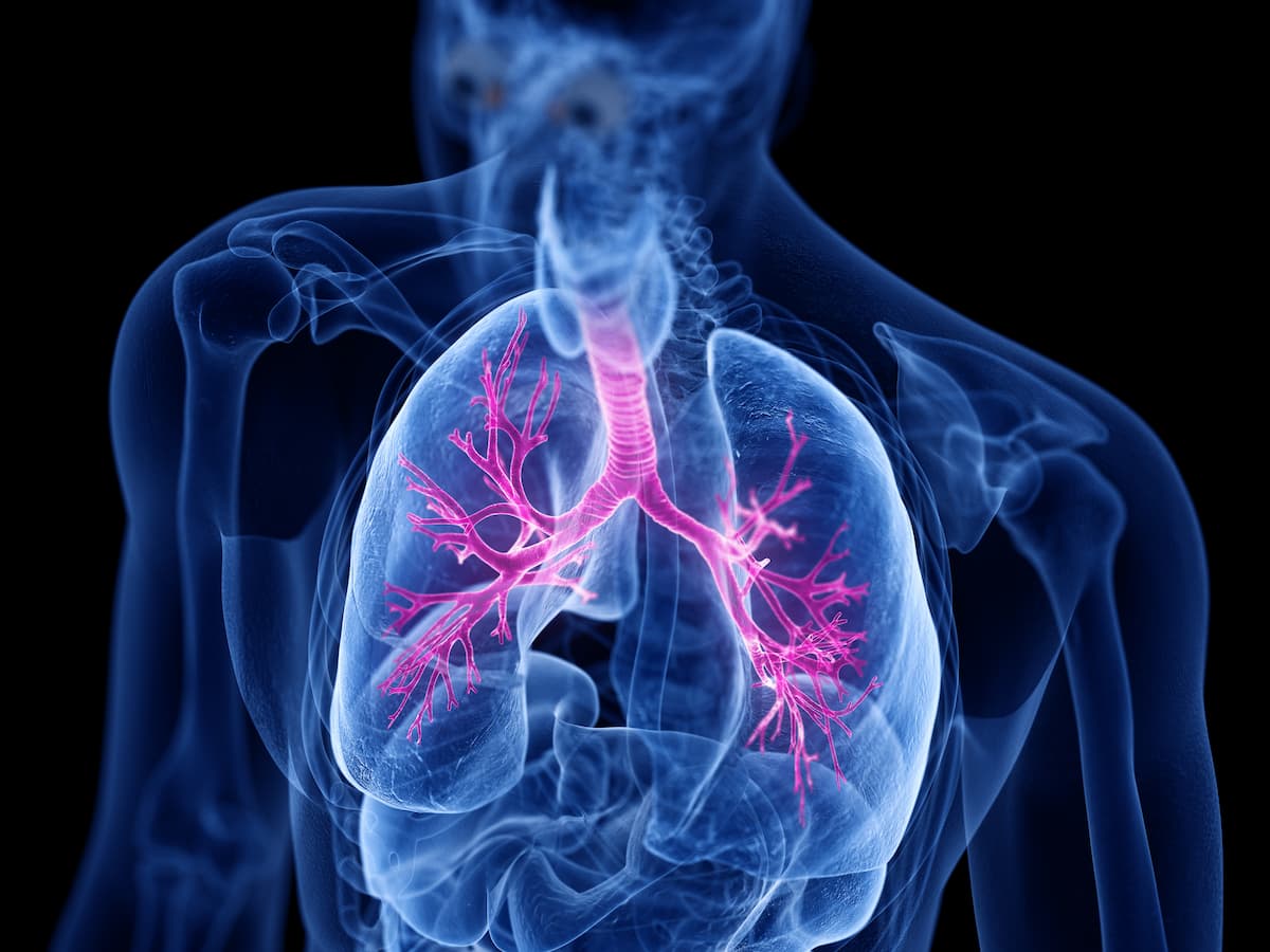
Atomic force microscopy analysis yielded distinct nanomechanical profiles in pulmonary fibrosis. | Image credit: Sebastian Kaulitzki - stock.adobe.com

Existing biomarkers to characterize pulmonary fibrosis are lacking in utility, they explained, leaving an “urgent need” for more reliable tools. The authors noted that regular histopathology, radiological data, and gene expression analysis are generally used to assess fibrotic tissues, and that nanomechanical properties are “usually overlooked” as potential biomarkers.
“Since PF progression involves changes in tissue structure and mechanics, mechanobiology plays a central role,” they said.
Advances in AFM technology have created the ability to assess mechanical properties at the cellular level, Stylianou and colleagues noted. The method has already been used for mechanical characterization of tissue biopsies.2
“However, most of these research efforts are mainly focusing on cancer, with limited studies in PF,” the authors explained.1
The investigators hypothesized that AFM might be able to identify unique nanomechanical fingerprints (NMFs) in tissue samples of patients with pulmonary fibrosis, and that those NMFs could serve as meaningful biomarkers to diagnose, stage, and track patients with the disease.
Stylianou and colleagues used AFM to assess biopsies taken from 5 patients with pulmonary fibrosis. Next, they used AMF in bleomycin-induced murine models of pulmonary fibrosis. They also studied the tool in mice treated with pirfenidone, which is known to reduce collagen type I.
The investigators found that AFM analysis yielded distinct nanomechanical profiles.
“Overall, fibrotic tissues showed markedly increased mechanical heterogeneity compared to non-fibrotic controls, reflecting the irregular and variable nature of ECM (extracellular matrix) remodeling in PF,” they said.
The investigators cautioned that their analysis is based on a small sample size, which they said was limited by a low availability of patients and by a limited number of patients willing to participate in the trial.
The authors found that not only could AFM identify unique nanomechanical features, but it could also detect subtle changes in nanomechanical properties as fibrosis progressed.
“Our results suggest that AFM-derived NMFs are sensitive enough to capture the mechanical alterations that occur during fibrosis progression, making them a promising tool for disease staging,” they wrote.
The investigators said their studies in mice suggest that these signatures might enable earlier diagnosis of the disease. Furthermore, they said nanomechanical changes might also be a useful means to monitor treatment efficacy. For example, they said AFM monitoring was able to show changes after mice were treated with pirfenidone.
Stylianou and colleagues said they do not see AFM as a full replacement for traditional histological or molecular assays but rather as a complementary method that can capture “often overlooked” biomechanical features of pulmonary fibrosis.
“The consistency between murine and human data underscores the potential use of NMFs as diagnostic or monitoring biomarkers in clinical settings,” they wrote.
They added that their findings suggest AFM could also have benefits in monitoring other diseases, potentially creating new pathways to personalized medicine.
References
1. Stylianou A, Polemidiotou K, Alimisis V, et al. Atomic force microscopy-based nanomechanical signatures for staging classification and drug response in pulmonary fibrosis. Small. Published online September 27, 2025. doi:10.1002/smll.202504526
2. Plodinec M, Loparic M, Monnier CA, et al. The nanomechanical signature of breast cancer. Nat Nanotechnol. 2012;7(11):757-765. doi:10.1038/nnano.2012.167
