
- Center on Health Equity & Access
- Clinical
- Health Care Cost
- Health Care Delivery
- Insurance
- Policy
- Technology
- Value-Based Care
Lung Ultrasound Can Accurately Detect ILD in Patients With RA
The meta-analysis of 9 studies showed high sensitivity with slightly lower specificity with the less-invasive option for detecting lung complications in patients with the autoimmune disease.
An ultrasound of the lungs is a reliable, radiation-sparing, and cost-effective option for detecting lung complications in patients with rheumatoid arthritis (RA), according to a compilation of available data.1 The meta-analysis, detailed in Rheumatology & Autoimmunity, showed that interstitial lung disease (ILD) was accurately detected in patients through lung ultrasound, regardless of symptomatic or asymptomatic disease.


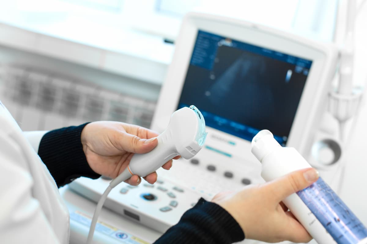
Ultrasound may be a more viable option than other methods, such as chest radiography and spirometry, to detect lung complications in rheumatoid arthritis. | Image credit: Kate - stock.adobe.com
The data suggest that lung ultrasound is a more viable option than other methods, such as chest radiography and spirometry. They also mirror those observed among patients with systemic sclerosis, citing a meta-analysis that showed a sensitivity rate of 94% and lower specificity of 64%.
Within RA, a previous study published in 2024 indicated that lung ultrasound was a useful detection method for ILD when used in combination with diffusing capacity of the lungs for carbon monoxide, though researchers of that study cautioned against substituting HRCT with the method. There were 192 patients included in this study, among whom researchers found that B-lines over 11.5 on the ROC curve were predictive of ILD.2
The current meta-analysis describes the findings from 845 patients across the 9 studies, which showed a high sensitivity rate (91%; 95% CI, 0.837-0.953) with slightly lower specificity (79.3%; 95% CI, 0.509-0.934). Area under the curve (AUC) was 0.926.
“Most studies in our meta-analysis used the detection of B-lines in LUS as the criterion for ILD diagnosis, although other sonographic features, such as pleural irregularity and subpleural nodules, have also been reported,” described the researchers. “B‐lines are vertical, laser‐like hyperechoic artifacts extending from the pleural line, indicative of pulmonary interstitial syndrome. These lines arise due to increased interstitial space or partial lung de-aeration, as seen in cases of pulmonary edema or ILD.”
Eight of the 9 studies used B-lines as a criterion for determining ILD, among which the researchers found a similar sensitivity rate of 90.6% (95% CI, 0.826-0.952) and specificity rate of 73.9% (95% CI, 0.439-0.911). The AUC was 0.916.
A smaller analysis included 5 studies that assessed B-lines in 14 LIS points. This subgroup analysis showed similar sensitivity and specificity rates, with a sensitivity rate of 89.1% (95% CI, 0.746-0.958) and specificity of 89.1% (95% CI, 0.612-0.977). AUC for these studies was 0.938.
Two of the studies assessed 72 LIS, which improved sensitivity (91.7%; 95% CI, 0.798-0.968) but reduced specificity (56.4%; 95% CI, 0.440-0.682).
“While scanning more LIS has demonstrated good sensitivity, it is time-consuming and may not be practical for daily clinical use. Simplified methods evaluating fewer LIS, targeting sites strongly associated with ILD, have shown acceptable accuracy,” wrote the researchers. “However, direct comparisons of LUS diagnostic accuracy based on the number of LIS examined are lacking, particularly in RA patients.”
Existing research has pointed to a positive correlation between the amount of B-lines on the ultrasound and high-resolution computed tomography scores, though the researchers of this latest analysis struggled to pinpoint the optimal cutoff for lung ultrasound.
The cutoff for the number of B-lines used to diagnose the lung complication varied across the 8 studies. Studies that used a threshold of 5 B-lines led to a comparable sensitivity rate (94.8%; 95% CI, 0.834-0.985) but lowered the specificity rate to 60.4% (95% CI, 0.140-0.935), while a threshold of 10 B-lines led to both reduced sensitivity (88.1%; 95% CI, 0.778-0.939) and specificity (54.3%; 95% CI, 0.452-0.631).
"In conclusion, our meta-analysis suggests that LUS is a valuable tool for detecting ILD in patients with RA, demonstrating high overall diagnostic accuracy," the authors wrote. "As a noninvasive, radiation-free imaging modality, LUS can be safely and repeatedly used to identify lung abnormalities in RA patients that may warrant further evaluation with HRCT, facilitating timely diagnosis and early intervention. However, there remains a need for a validated scoring system and standardized protocols for LUS application in assessing RA-ILD."
References
- Hidayat R, Audrey J, Tandaju JR, Fauzia F. Lung ultrasonography as a tool to detect interstitial lung disease in rheumatoid arthritis: A meta-analysis of diagnostic test accuracy studies. Rheumatol Autoimmun. Published online May 5, 2025. doi:10.1002/rai2.70008
- Santos-Moreno P, Linares-Contreras MF, Rodríguez-Vargas G-S, et al. Usefulness of lung ultrasound as a method for early diagnosis of interstitial lung disease in patients with rheumatoid arthritis. Open Access Rheumatol. 2024;16(9):9-20. doi:10.2147/OARRR.S441720


