- Center on Health Equity & Access
- Clinical
- Health Care Cost
- Health Care Delivery
- Insurance
- Policy
- Technology
- Value-Based Care
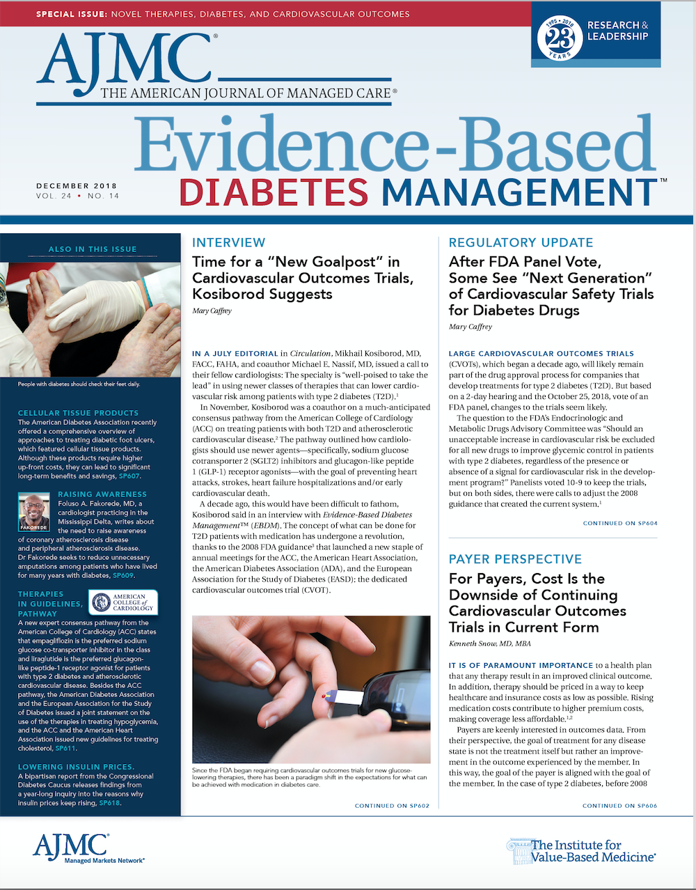
Clinical Evidence for and Cost-Effectiveness of Advanced Cellular Tissue Products for the Treatment of Diabetic Foot Ulcers
An estimated 30 million Americans are living with diabetes. Additionally, 84 million have prediabetes, a condition that will result in type 2 diabetes within 5 years if not properly treated. Long regarded as one of the most prevalent chronic diseases in United States, diabetes is also a leading cause of disability and the seventh-leading cause of death. Less discussed is one of the most common complications of diabetes: diabetic foot ulcers. If not properly treated with standard and adjunctive care, these chronic wounds can lead to permanent disability and premature death.
An estimated 30 million Americans are living with diabetes. Additionally, 84 million have prediabetes, a condition that will result in type 2 diabetes within 5 years if not properly treated.1 Long regarded as one of the most prevalent chronic diseases in United States, diabetes is also a leading cause of disability and the seventh-leading cause of death.2 Less discussed is one of the most common complications of diabetes: diabetic foot ulcers (DFUs). If not properly treated with standard and adjunctive care, these chronic wounds can lead to permanent disability and premature death.3 Although DFUs are not an inevitable comorbidity of diabetes, it has been estimated that the annual risk of developing a DFU may be as high as 4% and the lifetime risk may be as high as 34%.4
Without prompt intervention and proper treatment, DFUs will not heal, can cause soft tissue and/or bone infection, and may eventually require amputation of the affected limb or appendage. Unfortunately, this is not uncommon, as 1 in 6 patients with a DFU will undergo an amputation—making DFUs the leading cause of nontraumatic amputations in the country.5 Given the prevalence and risks associated with DFUs, it is imperative that clinicians understand, have access to, and use the best available science for the treatment of these hard- to-heal wounds.
In its recently released research compendium Diagnosis and Management of Diabetic Foot Complications, the American Diabetes Association (ADA) provides a comprehensive overview of the latest approaches for the manage- ment and treatment of DFUs and their complications. Produced by leading international DFU authorities, the compendium includes information about the use of adjunctive therapies, such as hyperbaric oxygen and negative pressure wound therapy, in instances in which DFUs do not respond to standard treatment.5 Among the treatments highlighted in the compendium that garnered the most attention were advanced cellular tissue products (CTPs). These are bioengineered cell-based therapies that supply the wound with the cells, tissues, proteins, and growth factors needed to support the healing process.
In addition to being supported by a wide body of clinical evidence demonstrating their effectiveness in facilitating wound healing, advanced CTPs have also been shown to generate significant cost savings for payers and the US healthcare system overall. This article will review the clinical evidence highlighted in the compendium demonstrating the effectiveness of CTPs in wound healing. It will also review recent literature supplemented by my own experience suggesting there are long-term cost benefits associated with using these therapies.
Advanced CTPs: Clinically Effective DFU Treatment
Patients with DFUs who are not responding to standard care are at great risk of not having their wounds heal and can suffer dire consequences. In practice, we clinicians see this reality daily as patients whose DFUs are not properly treated face increased risk of limb amputation, which results in higher mortality rates. Despite the availability of new therapies, DFUs remain notoriously difficult to treat. As most patients first experience a loss of feeling in the foot due to neuropathy, it is common for patients and providers to fail to notice a DFU until weeks after it has developed. Therefore, it is often recommended that clinicians be aware that advanced care may be required to ensure complete healing.6 In my practice, the use of advanced therapies has undoubtedly saved many limbs and lives.
Though treatment approaches for DFUs vary according to wound severity, clinicians commonly use an intensive adjunctive therapy in instances in which standard treatment—which typically consists of debridement, infection control, off-loading, and appropriate dressing—is not sufficient to reduce wound size over the first few weeks of care and/or close the wound in a timely fashion. In recent years, several innovative, advanced therapies have been developed for the treatment of DFUs, presenting new opportunities to better treat patients and minimize common risky and costly complications.
The compendium highlights 2 types of advanced, bioengineered CTPs used in adjunctive treatment of DFUs: an allogeneic bilayered human skin equivalent (HSE) and a dermal skin substitute (DSS). Unlike other types of CTPs, these are unique because they are cellular constructs and are both FDA-approved class III medical devices indicated specifically for the treatment of DFUs. To date, these are the only such therapies in their product class approved for this purpose.
Like human skin, HSE consists of 2 layers, has living cells, and contains structural proteins. The underside, or dermal, layer comprises a protein matrix of bovine type 1 collagen and neonatal human fibroblasts, while the epidermal layer consists of a stratified epithelium containing neonatal keratinocytes. Other components normally found in human skin—such as melanocytes, Langerhans cells, macrophages, and lymphocytes, as well as structures such as blood vessels, hair follicles, and sweat glands—are absent.
Two randomized controlled trials cited in the compendium have confirmed that patients treated with HSE showed significantly higher rates of healing and a shorter time to full wound closure than patients receiving standard care, making HSE among the best studied of all CTP therapies.7,8 The results of these rigorous trials led to HSE’s premarket approval by the FDA. Results from other studies not included in the compendium show HSE to be effective compared with other types of therapies. For example, a comparison study in a real-world setting found that treatment with an HSE increased the probability of healing by 97% compared with treatment with a dehydrated human amnion/chorion membrane, suggesting added benefit or that it was used more appropriately in practice.9
The dermal cellular CTP, DSS, is a single-layered construct of neonatal fibroblasts grown on absorbable mesh scaffold. The DSS delivers metabolically active human fibroblasts to a wound via a bioabsorable polyglactin mesh scaffold. Applying DSS imparts the benefits of human collagen, extracellular matrix proteins, and cytokines and other growth factors necessary for healing.
Among the studies cited by the ADA compendium, one large trial found that among patients with DFUs that have been present for more than 6 weeks, weekly application of the DSS resulted in significantly higher healing rates. Furthermore, treated patients were nearly twice as likely to have complete wound closure than those not treated with the DSS.10 Results of additional studies of patients treated with the DSS corroborate these findings. A study of patients with DFUs treated with the DSS found that 30% had achieved complete wound closure by week 12 compared with only 18.3% in the control group.11 Wounds treated with DSS—compared with other CTPs—were more likely to experience complete wound closure by week 12 (55% vs 32%) and week 24 (76% vs 50%) and had a significantly shorter mean time to full wound closure, evidence that DSS is perhaps among the most effective CTPs to date.12
Advanced CTPs: A Cost-Effective Solution
The cost of DFU treatment to the US health system is shockingly high—as much as $15 billion by some estimates.13,14 Another report found that treatment of DFUs may account for at least 33% of the direct medical costs associated with diabetes mellitus and that the cost to care for patients with DFUs is 5.4 times higher than the cost of care for those without them.15 The vast majority of these costs are related to patients with DFUs incurring higher emergency medical costs and being more likely overall to be admitted to the hospital. DFUs and related complications are one of the major reasons for hospitaliza- tions among patients with diabetes.16-18
Considering that foot ulcer reoccurrence is quite common and roughly 50% of all patients have a reoccurrence within 1 year after ulcer healing,5 it is important for clinicians to be equipped with the means to most effectively treat DFUs to help minimize hospitalizations, infections, and other associated complications that contribute to long-term costs.
Results from research have shown that despite the substantially higher upfront cost, advanced care that includes CTPs leads to long-term cost savings in the context of total cost of care related to DFUs. A meta-analysis of several dozen economic evaluations of cell-based tissue products found that CTPs resulted in shorter wound treatment periods, which, in turn, led to fewer complications and inpatient episodes.19 In other words, the more quickly a DFU progresses to closure, the lower the risk is of infection or other complications that can stall wound healing and result in costly surgeries and hospitalizations.
A more recent analysis that exclusively examined patients treated with cellular CTPs found that although patients received more intensive physician office and outpatient care, those costs were more than offset by reductions in lower-limb amputations and hospitalizations.20 The investigators in that study examined administrative claims data from Medicare beneficiaries between 2006 and 2012 and found that patients treated with CTPs had significantly lower amputation rates, fewer days hospitalized, and fewer emergency department visits than the control group, who received only standard care. During the 18-month follow-up period after treatment, average per-patient costs for treatment with an HSE were $5253 lower than those of the matched control. Patients treated with a DSS had costs $6991 lower than those of the control. These findings appear to be consistent with previous research showing reductions in lower-limb amputations and other resource-intensive healthcare procedures.21-22
In addition to improving healing, treatment of non—self-healing wounds with the products described above has the potential to lead to significant cost savings by reducing DFU-related complications such as osteomyelitis and amputation. An individual practitioner with a busy practice may reduce overall costs by tens or hundreds of thousands of dollars annually by using an advanced CTP in situations in which wound healing has not progressed using standard treatment.
Conclusion
Despite recent progress in the treatment of DFUs, these wounds remain a significant public health problem that will continue to require rigorous and evidence-based clinical practices. Proper use of evidence-based advanced adjunctive therapy is warranted. Advanced CTPs have shown promising scientific results for the treatment of DFUs,
in terms of both their clinical efficacy and the cost savings associated with shorter healing times and reduced risk of infection and related acute episodes and surgeries.
As clinicians, health systems, payers, and patient advocates grapple with how best to address this public health crisis, they would do well to consider the full suite of therapeutic options available and to use advanced interventions, when appropriate, that are clinically proved to help save limbs and lives.ABOUT THE AUTHOR:
Robert S. Kirsner, MD, PhD, is chairman of and holds the endowed Harvey Blank Chair in Dermatology in the Department of Dermatology and Cutaneous Surgery at the University of Miami Miller School of Medicine. He currently serves as director of the University of Miami Hospital and Clinics Wound Center and chief of Dermatology at the University of Miami Hospital and Jackson Memorial Hospital.
DISCLOSURES:
Dr Kirsner was a contributing author to Diagnosis and Management of Diabetic Foot Complications.
ACKNOWLEDGEMENTS:
The author would like to acknowledge Schmidt Public Affairs of Alexandria, Virginia, for editorial support provided.REFERENCES:
- CDC. National diabetes statistics report, 2017: estimates of diabetes and its burden in the United States. CDC website. cdc.gov/diabetes/ data/statistics-report/index.html. Published May 6, 2017. Accessed November 8, 2018.
- Kochanek KD, Murphy SL, Xu J, Tejada-Vera B. Deaths: final data for 2014. Natl Vital Stat Rep. 2016;65(4):1-122.
- Singer AJ, Tassiopoulos A, Kirsner RS. Evaluation and management of lower-extremity ulcers. N Engl J Med. 2017;377(16):1559-1567. doi: 10.1056/NEJMra1615243.
- Armstrong DG, Boulton AJM, Bus SA. Diabetic foot ulcers and their recurrence. N Engl J Med. 2017;376(24):2367-2375. doi: 10.1056/NEJMra1615439.
- Boulton AJM, Armstrong DG, Kirsner RS, et al. Diagnosis and Management of Diabetic Foot Complications. Arlington, VA: American Diabetes Association; 2018. professional.diabetes.org/sites/professional.diabetes.org/files/media/foot_complications_monograph.pdf. Published 2018. Accessed November 8, 2018.
- Kirsner RS, Bell D, Gibbons G, Ennis B, Masturzo A, Rothenberg G. Expert recommendations for optimizing outcomes utilizing Apligraf for diabetic foot ulcers. Podiatry Today website.podiatrytoday.com/files/ PT_orgo.pdf. Published 2012. Accessed November 8, 2018.
- Veves A, Falanga V, Armstrong DG, Sabolinski ML; Apligraf Diabetic Foot Ulcer Study. Graftskin, a human skin equivalent, is effective in the management of noninfected neuropathic diabetic foot ulcers: a prospective randomized multicenter clinical trial. Diabetes Care. 2001;24(2):290-295.
- Edmonds M; European and Australian Apilgraf Diabetic Foot Ulcer Study Group. Apligraf in the treatment of neuropathic diabetic foot ulcers. Int J Low Extrem Wounds 2009;8(1):11-18. doi: 10.1177/1534734609331597.
- Kirsner RS, Sabolinski ML, Parsons NB, Skornicki M, Marston WA. Comparative effectiveness of a bioengineered living cellular construct vs. a dehydrated human amniotic membrane allograft for the treatment of diabetic foot ulcers in a real world setting. Wound Repair Regen. 2015;23(5):737-744. doi: 10.1111/wrr.12332.
- Marston W, Hanft J, Norwood P, Pollak R; Dermagraft Diabetic Foot Ulcer Study Group. The efficacy and safety of Dermagraft in improving the healing of chronic diabetic foot ulcers: results of a prospective randomized trial. Diabetes Care. 2003;26(6):1701-1705.
- Warriner RA III, Cardinal M; TIDE Investigators. Human fibroblast derived dermal substitute: results from a treatment investigational device exemption (TIDE) study in diabetic foot ulcers. Adv Skin Wound Care. 2011;24(7):306-311. doi: 10.1097/01.ASW.0000399647.80210.61.
- Kraus I, Sabolinski ML, Skornicki M, Parsons NB. The comparative effectiveness of a human fibroblast dermal substitute versus a dehydrated human amnion/chorion membrane allograft for the treatment of diabetic foot ulcers in a real-world setting. Wounds. 2017;29(5):125-132.
- Rice JB, Desai U, et al. Healthcare resource utilization and economic burden of diabetic foot ulcers in privately-insured and Medicare populations. Paper presented at: SAWC Spring Conference; May 1-5, 2013. Denver, CO.
- Rice JB, Desai U, Cummings AK, et al. Burden of diabetic foot ulcers for Medicare and private insurers. Diabetes Care. 2014;37(3):651-658. doi: 10.2337/dc13-2176.
- Driver VR, Fabbi M, Lavery LA, Gibbons G. The costs of diabetic foot: the economic case for the limb salvage team. J Vasc Surg. 2010;52(suppl 3):17S-22S: doi: 10.1016/j.jvs.2010.06.003.
- Frykberg RG, Zgonis T, Armstrong DG, et al; American College of Foot and Ankle Surgeons. Diabetic foot disorders: a clinical practice guideline (2006 revision). J Foot Ankle Surg. 2006;45(suppl 5):S1-S66. doi: 10.1016/ S1067-2516(07)60001-5.
- Skrepnek GH, Mills JL, Lavery LA, Armstrong DG. Health care service and outcomes among an estimated 6.7 million ambulatory care diabetic foot cases in the US. Diabetes Care. 2017;40(7):936-942. doi: 10.2337/dc16-2189.
- Skrepnek GH, Mills JL Sr, Armstrong DG. A diabetic emergency one million feet long: disparities and burdens of illness among diabetic foot ulcer cases within emergency departments in the United States, 2006- 2010. PLoS One. 2015;10(8):e0134914. doi: 10.1371/journal.pone.0134914.
- Langer A, Rogowski W. Systematic review of economic evaluations of human cell-derived wound care products for the treatment of venous leg and diabetic foot ulcers. BMC Health Services Research. 2009;9:115. doi: 10.1186/1472-6963-9-115.
- Rice JB, Desai U, Ristovska L, et al. Economic outcomes among Medicare patients receiving bioengineered cellular technologies for treatment of diabetic foot ulcers. J Med Econ. 2015;18(8):586-595. doi: 10.3111/13696998.2015.1031793.
- Frykberg RG, Marston WA, Cardinal M. The incidence of lower-extremity amputation and bone resection in diabetic foot ulcer patients treated with a human fibroblast-derived dermal substitute. Adv Skin Wound Care. 2015;28(1):17-20. doi: 10.1097/01.ASW.0000456630.12766.e9.
- Redekop WK, McDonnell J, Verboom P, Lovas K, Kalo Z. The cost effectiveness of Apligraf treatment of diabetic foot ulcers. Pharmacoeconomics 2003;21(16):1171-1183. doi: 10.2165/00019053-200321160-00003.

Quality of Life: The Pending Outcome in Idiopathic Pulmonary Fibrosis
February 6th 2026Because evidence gaps in idiopathic pulmonary fibrosis research hinder demonstration of antifibrotic therapies’ impact on patient quality of life (QOL), integrating validated health-related QOL measures into trials is urgently needed.
Read More
What It Takes to Improve Guideline-Based Heart Failure Care With Ty J. Gluckman, MD
August 5th 2025Explore innovative strategies to enhance heart failure treatment through guideline-directed medical therapy, remote monitoring, and artificial intelligence–driven solutions for better patient outcomes.
Listen
Building Trust: Public Priorities for Health Care AI Labeling
January 27th 2026A Michigan-based deliberative study found strong public support for patient-informed artificial intelligence (AI) labeling in health care, emphasizing transparency, privacy, equity, and safety to build trust.
Read More

