- Center on Health Equity & Access
- Clinical
- Health Care Cost
- Health Care Delivery
- Insurance
- Policy
- Technology
- Value-Based Care
Patients With AGA Treated With PRP Display Hair Regrowth, Potential Microbiome Rebalance
Platelet-rich plasma (PRP) treatment increased hair regrowth in patients with androgenetic alopecia (AGA) and may help rebalance the scalp microbiome, although the link between the microbiome changes and hair growth needs future investigations.
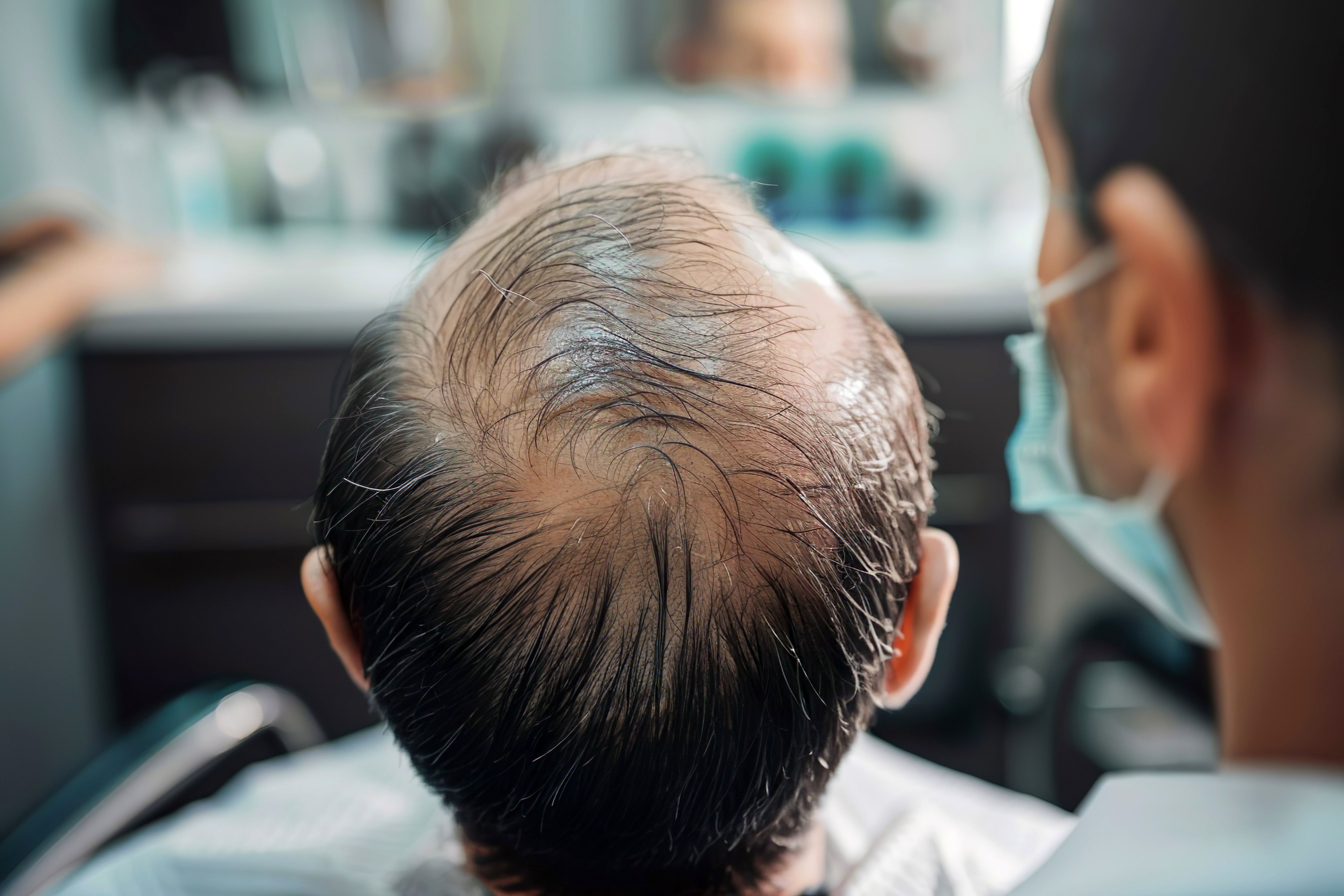
Male pattern baldness | Image Credit: Oksana - stock.adobe.com

Patients with androgenetic alopecia (AGA) found platelet-rich plasma (PRP) treatment led to increased hair growth and showed potential in scalp microbiome rebalancing.
A small sample study was performed on 14 participants with AGA to analyze the clinical efficacy while observing results before treatment (TB) and after treatment (TA).1
The study focused on scalp microbiome because research points to the imbalance in microbiota as a trigger for AGA, while the most common form of bacteria found in scalp swabs of healthy individuals are Cutibacterium spp. and Staphylococcus spp., accounting for approximately 90% of the total gene sequence.
Currently there are only 2 drugs approved by the US FDA for AGA treatment, oral finasteride and topical minoxidil, both of which come with serious adverse effects and are oftentimes not effective among patients. PRP treatment has been a successful method in treating AGA, proving so through basic research and clinical trials over the past decade.
Research has suggested an interplay between the gut and skin microbiome in relation to hair biology.2 While it is still unknown how the gut microbiota may influence AA pathogenesis, the association of AA and gut dysbiosis diseases is prominent as seen in inflammatory bowel disease, ulcerative colitis, and Crohn disease.
Prior to treatment, patients had 36 mL of peripheral blood extracted from each AGA volunteer.1 Then, PRP was categorized into 3 parts, prefrozen at –80 °C for a 24 hour period. Afterwards, PRP was freeze-dried using a freeze-drying machine for 48 hours. Participants were treated once a month and blood samples were collected after 3 months to isolate the PRP. Overall, treatment took place over a 6-month duration.
Clinical efficacy of PRP in AGA was evaluated through photography and dermascopy. Swab sampling was conducted using the scalp area of the patients with AGA. Following procedure, every swab was put into a 50 mL centrifuge tube and stored at –80 °C until the DNA was extracted from the sample.
Patients with AGA showed an increased level of macroscopic hair regrowth following 6 treatments with PRP. Dermatoscopes showed significant changes in a multitude of parameters in hair growth. Before treatment, 14 patients with AGA were sampled and after treatment, 8 samples were collected. Bacterial population was slightly higher in the TA group compared with the TB group but there were no statistically significant differences between the 2 groups.
While there were differences among the TA and TB groups, there were no major differences observed (P = .203).
Looking at the major types of bacteria, like phylum, the TB group was dominated by Firmicutes (39.4%), followed by Proteobacteria (23.4%) and Actinobacteriota (21.5%).
In the bacterial communities like genera, both the TB and TA groups had high levels of Staphylococcus (TB: 28.3%, TA: 21.7%), Lawsonella (TB: 10%, TA: 6.7%), Cutibacterium (TB: 8.5%, TA: 13.9%), and Bacteroides (TB: 5.1%, TA: 4.1%).
When examining individual bacterial species, uncultured_bacterium was the most abundant in both groups (TB: 39.1%, TA: 41.5%). This was followed by uncultured Staphylococcus (TB: 12.2%, TA: 7.4%) and Staphylococcus warneri (TB: 7.4%, TA: 2.4%).
The scalp microbial composition of the scalp before and after treatment expressed discernible differences at the species and genus levels.
Throughout the TB group, 3 bacterial genera were abundant, Brevundimonas, uncultured, and Eremococcus. There were 14 genera overall that were enriched in the TA group, known as Paenibacillus, Aeribacillus, Spirosoma, Comamonas, Cereibacter, Massilia, Nocardioides, Hydrogenophilus, Granulicatella, Geobacillus, Meiothermus, Pseudomonas, Thermus, and Brevibacillus.
Data found Pseudomonas_sp. was more abundant in the TA group (2.05%) compared with the TB group (0.68%).
The study highlighted the distinct role microbial ecological imbalance plays in scalp diseases because the skin hosts a diverse community of microbes. Following treatment for AGA, hair density grew instantly, scalp composition improved, and the ratio of g_Cutibacterium and g_Staphylococcus were normal. Additionally, Lawsonella was elevated in the TB group compared with the TA group for the first time in scalp microbiota.
Similarly, a 2-sample Mendelian randomization study suggested that certain types of bacteria, like Olsenella, Ruminococcaceae UCG-004, and Ruminococcaceae UCG-010, might increase the risk of AGA.3 On the other hand, bacteria belonging to Ruminiclostridium 9, Anaerofilum, and the Acidaminococcaceae family seemed to have a protective effect. These findings suggest a connection between specific gut microbes and AGA, opening the door for developing more targeted probiotics to potentially help manage hair loss.
While PRP treatment does show signs of positive impacts on the composition of scalp microbiota, further research is necessary to investigate the impact microbial communities have on patients with AGA to determine whether PRP has a therapeutic role after altering the composition of the scalp.
References
1. Zhang Q, Wang Y, Ran C, et al. Characterization of distinct microbiota associated with androgenetic alopecia patients treated and untreated with platelet-rich plasma (PRP). Animal Model Exp Med. 2024;7(2):106-113. doi:10.1002/ame2.12414
2. Carrington AE, Maloh J, Nong Y, Agbai ON, Bodemer AA, Sivamani RK. The gut and skin microbiome in alopecia: associations and interventions. J Clin Aesthet Dermatol. 2023;16(10):59-64.
3. Fu H, Xu T, Zhao W, Jiang L, Shan S. Roles of gut microbiota in androgenetic alopecia: insights from Mendelian randomization analysis. Front Microbiol. 2024;15. doi:10.3389/fmicb.2024.1360445
The Breakdown: Breast Cancer Research Awareness Day
August 19th 2025Breast cancer is the second most common cancer among women and the second leading cause of cancer-related deaths among women in the US. In light of Breast Cancer Research Awareness Day, The American Journal of Managed Care® breaks down the most recent advancements in breast cancer prevention, screening, and therapies.
Listen
