- Center on Health Equity & Access
- Clinical
- Health Care Cost
- Health Care Delivery
- Insurance
- Policy
- Technology
- Value-Based Care
Nanoparticle Delivery Holds Promise for Treatment of PAH
Aerosolized vascular endothelial growth factor/stromal cell-derived factor-1α (VEGFNP/SDFNP) led to a lower pulmonary arterial pressure and prevented right ventricular hypertrophy after monocrotaline (MCT) injection in rats.
Vascular endothelial growth factor/stromal cell-derived factor-1α (VEGFNP/SDFNP) delivery significantly reduced the pulmonary arterial pressure, pulmonary vascular resistance, and thickening of the distal pulmonary vessels in rats with pulmonary arterial hypertension (PAH), according to a study in Pulmonary Circulation.1
“Right ventricular hypertrophy was nearly prevented in the rats, indicating that the VEGFNP/SDFNP treatment had profoundly delayed the progress of PAH development,” the authors wrote.
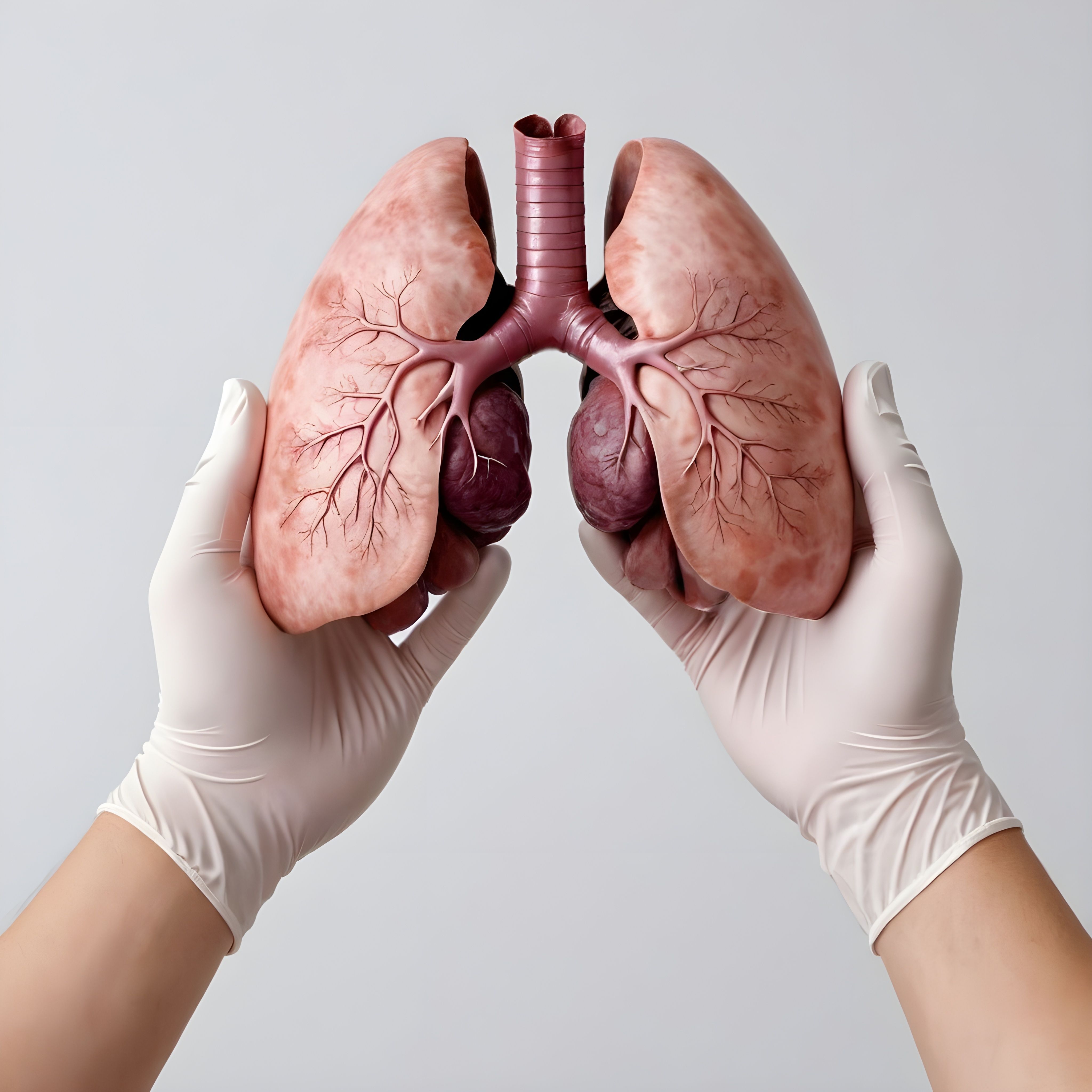
Lung analysis concept | image credit: fauzi - stock.adobe.com

Using Dulbecco's phospate-buffered saline (PBS), nanoparticles consisting of 4 µg SDF (SDFNP) and 8 µg VEGF (VEGFNP) were diluted together to a volume of 0.25 mL and were subsequently aerosolized into rat lungs on Day 14, at an early- and mid-stage of disease, after monocrotaline (MCT) injection. Analyses were performed on Day 28.
Gene expression analysis of MCT lungs was like that of the endothelial cell markers, VE-cadherin, KDR, and BMPR2, in the MCT + VEGFNP/SDFNP treated lungs. Both were significantly lower than no treatment control. These findings caused the researchers to reject an earlier assumption that the therapeutic effect of VEGFNP/SDFNP must be related to the endothelial cell growth function of VEGF. Researchers hypothesized that VEGFNP/SDFNP could help repair existing endothelium by replacing dysfunctional or apoptotic cells.
VEGFNP/SDFNP significantly increased the endothelial cell marker eNOS and decreased α-SMA expression in the MCT-treated lungs. VEGFNP/SDFNP delivery did not change the reduced expression of VE-cadherin, suggesting that angiogenesis did not influence the recovery of the lost endothelial cells with the treatment, the study found.
The mean right ventricular systolic pressure, pulmonary vascular resistance indices, and the Fulton indices [−RV/(LV + Septum)] of the treatment groups were as follows: control (29 mmHg, 0.6, 0.22), MCT (70 mmHg, 3.2, 0.44), MCT + VEGFNP/SDFNP (40 mmHg, 1.7, 0.23).
VEGFNP/SDFNP delivery prolonged the thickening of distal pulmonary vessels. MCT resulted in 46±12 almost occluded vessels in a whole lung section, and the same figure was 2±3 for MCT + VEGFNP/SDFNP.
VEGFNP/SDFNP delivery also significantly lowered the right ventricular systolic pressure (RVSP) and the pulmonary vascular resistance (PVR) in rats treated with MCT. Mean RSVP of the control, MCT and MCT plus VEGFNP/SDFNP groups were 31, 81 and 54. The mean PVR indices of the groups were 0.39, 2.33, and 1.51 mm Hg/mL/min.
The structural/receptor endothelial cell markers of the MCT and the control groups were similar, suggesting a relative increase in eNOS expression in the MCT lungs. Expression of structural/receptor endothelial cell markers differed from that of eNOS.
VEGFNP/SDFNP treatment reduced medial thickening of distal pulmonary vessels caused by MCT. But there was a mean of 43 initially muscularized, distal thin0walled vessels throughout VEGFNP/SDFNP-treated lungs, compared with means of 2 and 57 for the control and MCT groups, respectively.
Numerous studies point to the protective effect of VEGF in stymying development of severe PAH, including delivery of VEGF to hypoxic rat lungs through adenovirus-mediated gene transfer. In chronic hypoxic rats and mice, blocking VEGF signaling by inhibition of its receptor (VEGFR2/KDR/flk-1) with SU5416 significantly worsened PAH. Deactivation of the endothelial VEGF receptor gene in mice resulted in similar effects compared with SU5416. It was discovered that some patients with heritable PAH carried heterozygous, mutated loss-of-function of the KDR gene.
CsA-treated Sprague Dawley rats and nude rats were used to prevent the rats’ immune systems from eliminating human proteins delivered to their lungs.
A previous study established that adenoviral-mediated VEGF overexpression in the lungs attenuates development of hypoxic pulmonary hypertension, in part by protecting endothelium-dependent function.2
“These findings differed from those in MCT-treated Sprague Dawley rat lungs, which often were overwhelmed with macrophages and perivascular inflammation. Since inflammation plays an important role in the development of PAH, lacking this factor may have, in part, accounted for the therapeutic effects of VEGFNP/SDFNP observed in this study,” the authors wrote.
References
- Guarino VA, Wertheim BM, Xiao W, et al. Nanoparticle delivery of VEGF and SDF-1α as an approach for treatment of pulmonary arterial hypertension. Pulm Circ. 2024;14:e12412. doi:10.1002/pul2.12412
- Partovian C, Adnot S, Raffestin B, et al. Adenovirus-mediated lung vascular endothelial growth factor overexpression protects against hypoxic pulmonary hypertension in rats. Am J Respir Cell Mol Biol. 2000; 23: 762–771. doi: 10.1165/ajrcmb.23.6.4106
