- Center on Health Equity & Access
- Clinical
- Health Care Cost
- Health Care Delivery
- Insurance
- Policy
- Technology
- Value-Based Care
Management Strategies for Metabolic Dysfunction–Associated Steatotic Liver Disease (MASLD)
Abstract
Metabolic dysfunction–associated steatotic liver disease (MASLD) is characterized by hepatic steatosis that is confirmed by imaging or histology in the setting of at least 1 metabolic risk factor in the absence of significant alcohol consumption. Nonalcoholic steatohepatitis, or NASH, was recently renamed metabolic dysfunction–associated steatohepatitis (MASH); it represents the progressive form of MASLD. MASH is defined by hepatic steatosis, lobular inflammation, and ballooning degeneration (hepatocellular injury) in a characteristic histologic pattern. Multiple pathophysiologic mechanisms underlie the development of MASLD, and multiple factors (eg, metabolic, hormonal, genetic, nutritional, and epigenetic components) are related to liver injury. MASH has a prevalence in the United States of 1% to 6%, and it is expected to rise in the next decade. Individuals living with MASH frequently suffer from comorbidities such as type 2 diabetes and cardiovascular disease. Several guidelines have been published to support the timely diagnosis of MASH that incorporate noninvasive tests that obviate the need for liver biopsy. Multiple MASH treatment options are in various stages of development. The THR-β agonist resmetirom, approved by FDA in March 2024, offers a liver-directed treatment for those patients living with moderate to severe fibrosis without cirrhosis. Considering the progressive nature of the disease and the availability of a treatment that can be initiated early to halt MASH progression, patients who have risk factors for MASH should urgently be encouraged to visit their health care providers for MASH screening.
Am J Manag Care. 2024;30(suppl 9):S159-S174. https://doi.org/10.37765/ajmc.2024.89635
For author information and disclosures, see end of text.
A Note on Nomenclature of Nonalcoholic Fatty Liver Disease and Nonalcoholic Steatohepatitis
The naming convention of nonalcoholic steatohepatitis (NASH) was first established in 19801; since then, several alternative names for NASH have been proposed. From 2020 to 2022, a comprehensive and inclusive Delphi consensus process led to the adoption of new steatotic liver disease nomenclature. Thus, the terms metabolic dysfunction–associated steatotic liver disease (MASLD) and metabolic dysfunction–associated steatohepatitis (MASH) are now used.2 Subsequent analyses have demonstrated that patients described with the new nomenclature are relatively indistinguishable from those classified using the former nonalcoholic fatty liver disease (NAFLD) nomenclature.2-4 Thus, the findings of studies reported over 3 decades that describe patient characteristics, treatment, and outcomes for NAFLD still apply when using the MASLD nomenclature. For these reasons, in this review, the terms MASLD and MASH will be used.
Background
MASLD is characterized by hepatic steatosis that is confirmed by imaging or histology in the setting of at least 1 metabolic risk factor in the absence of significant alcohol consumption (Figure 1).5-11 Disease severity within MASLD covers a spectrum from MASL with little or no inflammation or hepatocyte injury to MASH, at-risk MASH (defined as MASH with significant fibrosis), or MASH cirrhosis.5-11 MASH represents the progressive form of MASLD; it is defined by hepatic steatosis, lobular inflammation, and ballooning degeneration (hepatocellular injury) in a characteristic histologic pattern.5-11
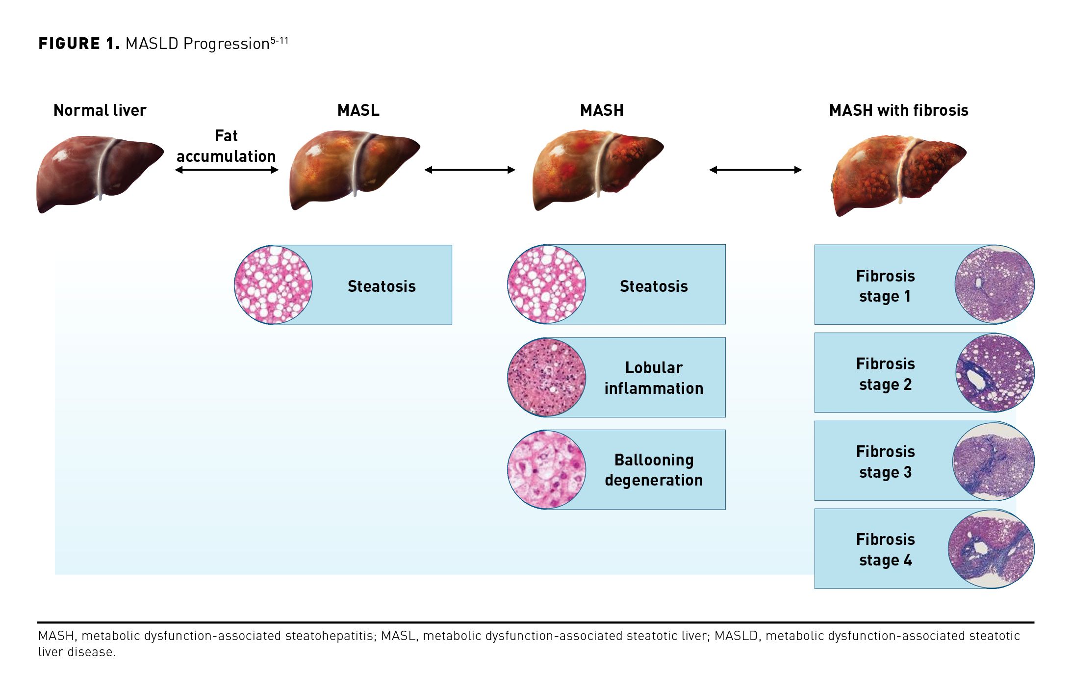
A formal diagnosis of MASH can only be confirmed through liver biopsy.5,7,12 However, this procedure is less commonly used in real-world clinical practice due to its invasive nature, procedure-related limitations, and potential for serious complications.13-20 As such, the diagnosis of suspected MASH in most patients is based upon clinical, laboratory, and imaging data that are collectively referred to as results of noninvasive tests (NITs) with appropriate exclusion of other liver conditions.21 Risk stratification among patients with MASH can be performed noninvasively according to patient characteristics such as the number of metabolic risk factors (eg, obesity, type 2 diabetes [T2D], hypertension, and dyslipidemia), age, platelet count, the results of various proprietary tests, and the output of algorithms.
Epidemiology of MASLD and MASH
MASLD is the most common chronic liver disease in the United States, affecting an estimated 31% of the nation’s adult population.22 The precise epidemiology of MASH—particularly the population of patients with clinically significant liver fibrosis (fibrosis stage 2/3 [F2/3])—is difficult to determine due to uncertainty in risk stratification, differences in diagnostic methods used to identify the condition (ie, liver biopsy or imaging- or biomarker-based NITs), and disparities in the definition of the disease.23,24
Harrison and colleagues prospectively assessed the prevalence and severity of MASLD and MASH among 664 asymptomatic middle-aged Americans.25 Patients were evaluated using results of NITs and liver biopsies to obtain point estimates of the prevalence and severity of MASLD/MASH. The prevalence of MASLD was 38% (95% CI, 34%-41%), and the prevalence of MASH was 14% (95% CI, 12%-17%). No patient had cirrhosis on biopsy; however, significant fibrosis (≥ F2) was present in 5.9% (95% CI, 4%-8%) of patients, and bridging fibrosis (F3) was revealed in 1.6% (95% CI, 1%-3%).25 However, the estimated prevalence of MASH in the United States (1%-6%) is projected to increase substantially over the next decade.26-33
MASH is more common in patients with T2D or metabolic syndrome. There may be an 18-fold higher risk of MASH among people with diabetes.29 For people living with obesity, the global prevalence of MASLD is 70% to 80% and rises to 90% for those living with morbid obesity. Of the 18.2 million people living in the United States with T2D and MASLD, 6.4 million individuals have MASH.30
Men may be slightly more likely to develop MASH than are women; however, women experience a late peak in MASH diagnosis postmenopause.32 Both MASLD and MASH are more common among Hispanic individuals than among those belonging to other ethnic groups; MASLD is diagnosed in 58% of Hispanic patients and 45% of White patients, and MASH is diagnosed in 19% and 10% of patients in these groups, respectively.31 The presence of the PNPLA3*G allele may contribute to the higher prevalence of advanced MASLD among Hispanic individuals.32
Pathophysiology and Natural History of MASLD and MASH
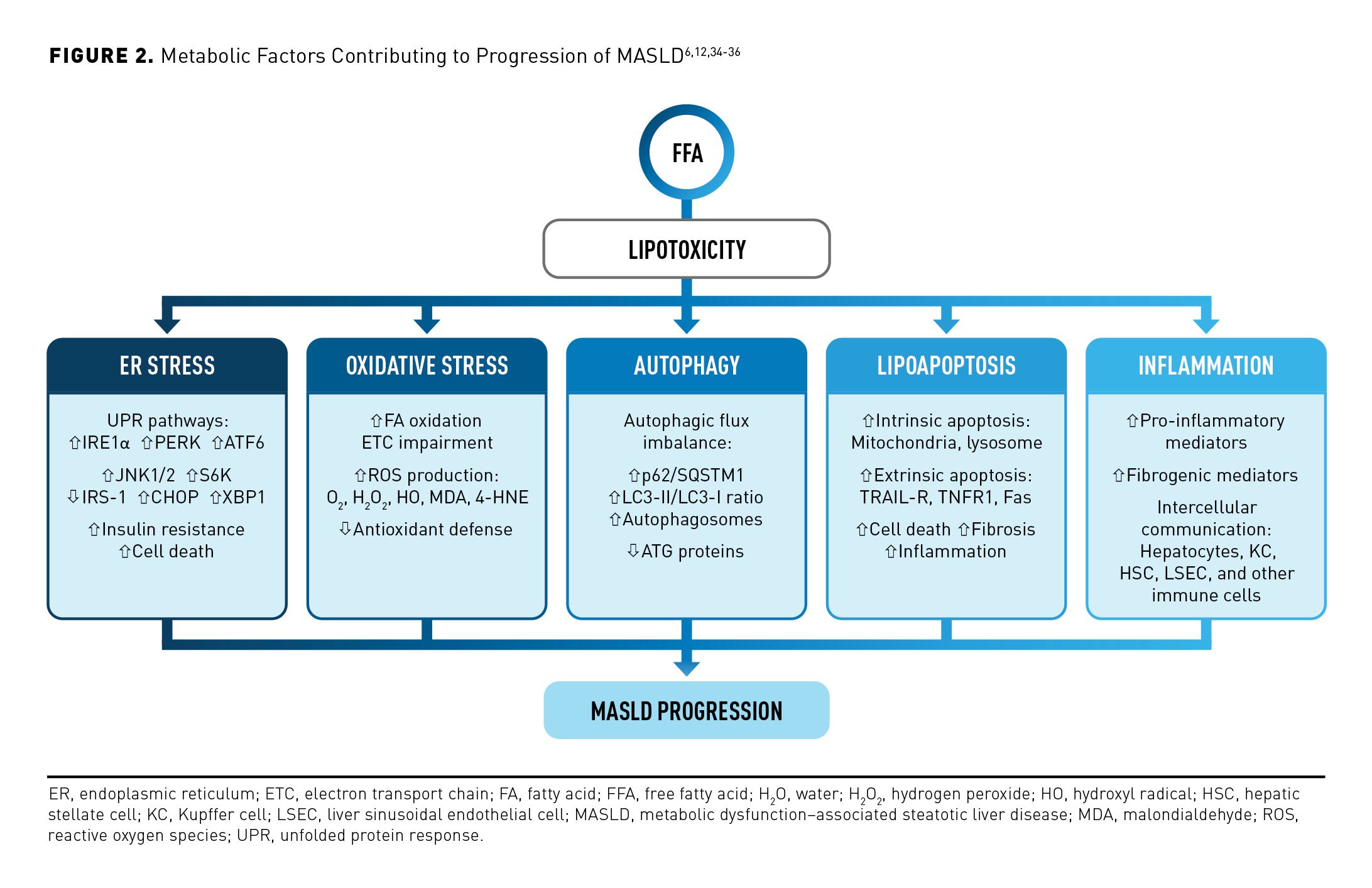
Multiple pathophysiologic mechanisms underlie the development of MASLD with multiple factors (eg, metabolic, hormonal, genetic, nutritional, and epigenetic components) related to liver injury.6,12,34 Hepatic fatty acid exposure and systemic insulin resistance resulting from dietary, genetic, and metabolic factors lead to hepatocyte lipotoxic injury and stress, which may contribute to mitochondrial dysfunction in MASLD.6,35 Impaired thyroid hormone signaling in hepatocytes worsens hepatic lipotoxicity and drives progression of MASLD to MASH with liver fibrosis.36 Mitochondrial dysfunction in the liver leads to the generation of reactive oxygen species and hepatocellular oxidative injury, which contribute to necroptosis and downstream inflammation.6,35 Adipocytes release nonesterified fatty acids as a result of impaired insulin responsiveness, leading to lipid accumulation in the liver. In MASLD, de novo lipogenesis is also increased, contributing to the oversupply of free fatty acids in hepatocytes. Together, the increased supply of exogenous and endogenous free fatty acids leads to lipotoxic stress.
Fatty acids can be eliminated by the formation of triglyceride and its export into the blood as very low–density lipoprotein or by mitochondrial β-oxidation. Thyroid hormone is a regulator of mitochondrial biogenesis and oxidative capacity; however, in the setting of MASLD, conversion of thyroxine (the inactive form of thyroid hormone) to triiodothyronine (T3) (the active form) is inadequate, and the MASLD liver is in a state of relative hypothyroidism. This can be counteracted by drugs that activate the thyroid hormone receptor (THR). The predominant THR in the liver is the β isoform; THR-β agonism results in an increase in fat metabolism through mitochondrial β-oxidation, a reduction in fatty acids and other lipotoxic lipids such as ceramides, and an increase in cholesterol clearance.
There is an increase in inflammatory cytokines and an accumulation of hepatic triglycerides as dysfunctional adipose tissue causes the release of nonesterified fatty acids.36 This perpetuates a cycle of oxidative stress, further inflammation, and eventual fibrogenesis.6 Endoplasmic reticulum (ER) stress is also a component of lipotoxic liver injury.35 Stress in the ER brings on changes in signaling to a repair response to reestablish homeostasis; however, prolonged ER stress reduces the ability of hepatocytes to restore homeostasis.35 Eventually, prolonged ER stress leads hepatocytes to undergo apoptosis and begins the wound-healing signaling cascade that is hallmarked by inflammation, vascular remodeling, fibrogenesis, and sequestering of immature hepatocytes.27 Kupffer cell cytokine production also contributes to the development of fibrosis in the liver, playing a central role in activating stellate cells to initiate and perpetuate fibrogenesis.37 Further, some individuals may be prone to the development of MASH due to genetic abnormalities, and they may not present with obesity and insulin resistance.6,37 The metabolic factors contributing to the progression of MASLD to MASH are described in Figure 2.6,12,34-36
Clinical Presentation and Diagnosis
MASLD represents a spectrum of liver diseases characterized by hepatic steatosis in the setting of at least 1 metabolic risk factor.5,6 Current guidelines from Europe, Japan, and the US define MASLD as the presence of hepatic steatosis (> 5% of hepatocytes show evidence of steatosis) confirmed by imaging or histology in the absence of significant alcohol consumption.5-11 Diagnosis of MASLD requires that excessive alcohol consumption is ruled out as a contributor of fibrosis.5 Levels of alcohol consumption considered to be excessive in this context are less than 30 g/d for men or less than 20 g/d for women.23
MASH is a hepatic disease, yet patients with MASLD/MASH typically present with obesity as defined by excessive body mass index (BMI) and/or other components of the metabolic syndrome.12 On assessing patients at risk, care providers should also look for physical indicators of advanced liver disease including hepatomegaly or splenomegaly (enlarged liver or spleen, respectively), ascites, muscle loss, or signs of insulin resistance such as acanthosis nigricans (ie, darkening of skin in folds and creases).5,12 Routine screening for MASLD based on the presence of metabolic factors or T2D is currently recommended by the World Gastroenterology Organisation (WGO); Italian, Japanese, United Kingdom, and European guidelines; and recommendations by the American Association of Clinical Endocrinology (AACE), American Association for the Study of Liver Diseases (AASLD), and the American Gastroenterological Association (AGA) (Table 1).7,8,23,24,38

The Central Role of Noninvasive Tests
Liver biopsy is the classic tool used in the diagnosis of MASLD/MASH, although it has limited practicality in routine clinical care due to its invasive nature, associated risks, and reader variability.39 Accordingly, the current guidance extensively incorporates the use of NITs as markers for identification, determination of severity, monitoring, and establishment of prognosis of patients with MASLD/MASH (Table 2).7,24,38,40-56
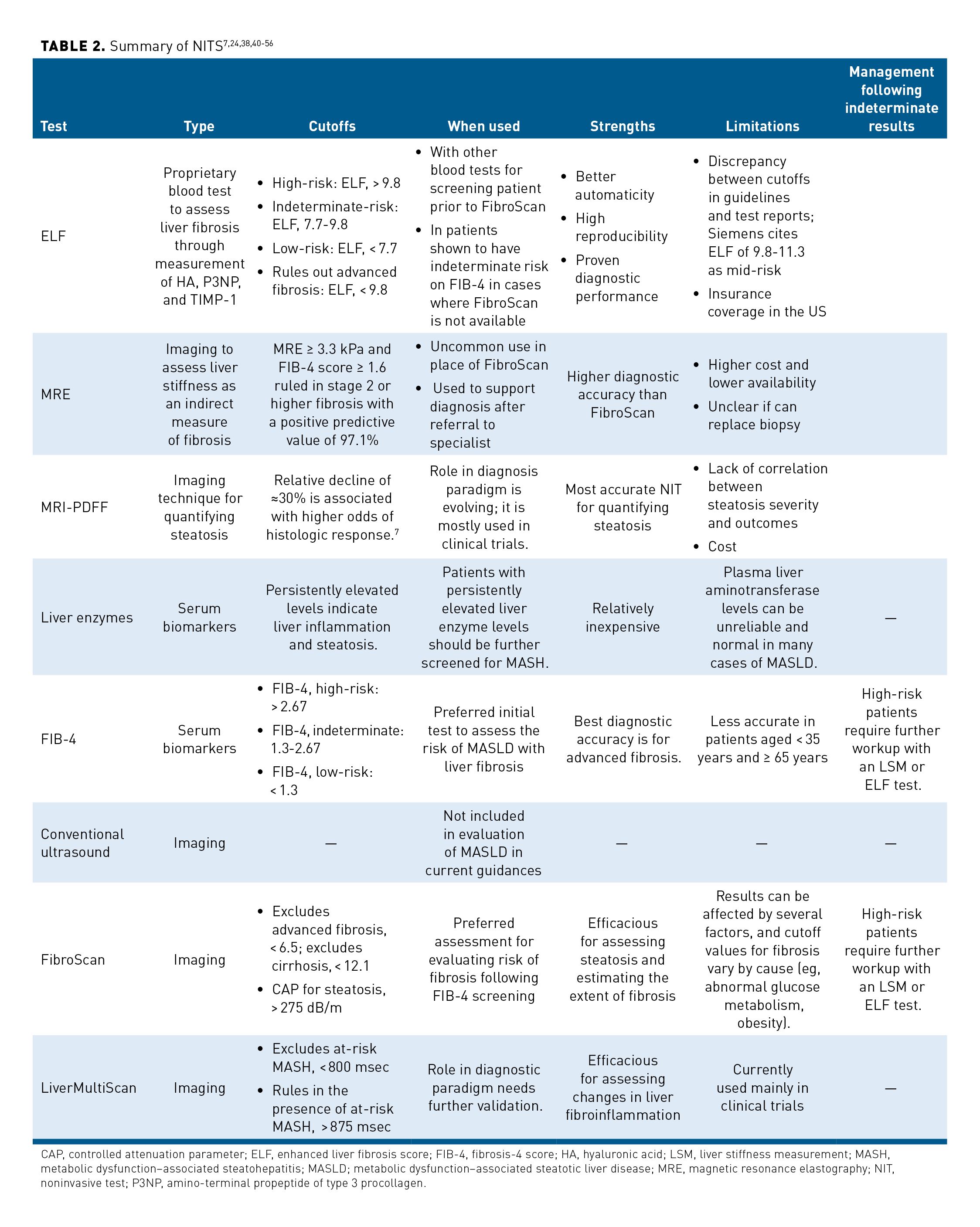
NITs include testing of blood-based biomarkers (the liver enzymes aspartate aminotransferase [AST], alanine aminotransferase [ALT], and γ-glutamyl transferase [GGT]).57 In addition, the enhanced liver fibrosis (ELF) test (Siemens Healthineers USA) is a proprietary blood test to assess liver fibrosis using measures of hyaluronic acid, P3NP, and TIMP-1.58 The ELF test has approval in the US for prognostic use; it has received FDA breakthrough device designation for diagnostic use,58 and it has high sensitivity and specificity for detecting clinically advanced liver fibrosis and good prognostic value in predicting liver-related events and mortality in patients with chronic liver disease.
Other tests in development and not yet available in the clinic include the ProC3 test, which is an enzyme-linked immunosorbent assay (ELISA) that measures the cleavage of propeptide collagen fragments released during active fibrogenesis.59 In addition, the NIS2+ (GENFIT) test uses 2 biomarkers (miR-34a-5p and YKL-40); it has been used more extensively in conjunction with clinical trials of MASH treatments.60 Further, the FIB-4 score is a relatively inexpensive, easy to use, and validated tool that employs age, platelet counts, and ALT and AST measurements as parameters to identify patients with advanced fibrosis; it is featured in European Association for the Study of the Liver (EASL), AACE, AASLD, and AGA guidelines/guidance for the diagnosis of NASH/MASH because of its ease of calculation and minimal cost.7,8,23,24 Its limitations include diagnostic cutoff ranges that may not be reliable in relevant populations (eg, patients with T2D),61 lack of accuracy in both younger (age, < 35 years) and elderly patients,40 and limited data for monitoring disease evolution and treatment response. It also depends upon variations in liver enzymes that may be attributable to other causes.
The most common imaging method for identifying patients with hepatic steatosis is conventional ultrasound, which is widely available, inexpensive, and well established. It has inherent limitations including poor sensitivity, overestimation of steatosis (especially in patients with obesity), imprecision in quantifying steatosis, and high operator variability and reproducibility.7,62,63 Because of these limitations, routine ultrasound examination is not part of the current diagnostic algorithms for patients with suspected MASLD.
FibroScan (Echosens) is a noninvasive elastography test that uses vibration-controlled transient elastography (VCTE) for liver stiffness measurement (LSM) as a surrogate for fibrosis and controlled attenuation parameter (CAP) to assess hepatic steatosis.64 Other ultrasound-based technologies for assessing liver stiffness and fibrosis staging include visual transient elastography, sound touch elastography, and sound touch quantification. The LSM performs similarly to biopsy in predicting liver-related events.65
After the FIB-4 screen, elastography is the preferred imaging method; it may be used alone or in conjunction with blood tests for increased diagnostic specificity.66 Drawbacks include decreased accuracy in severely obese individuals, limited insurance coverage and data for monitoring patients with MASLD, and the lack of discrete cutoffs for individual fibrosis stages. Magnetic resonance elastography (MRE) uses MRI and a low-frequency mechanical vibrating pad to assess liver stiffness as an indirect measure of fibrosis.41,67 LSM by MRE may be used alone or in combination with blood tests for increased diagnostic specificity.42,43
MRE is considered the most accurate measure of LSM in MASLD.44 Current guidelines recommend considering MRE when LSM by VCTE/FibroScan and FIB-4 are indeterminate due to relatively higher cost and lower availability as compared with those of ultrasound-based technologies. Currently, there are at least 650 machines in the US, and 1200 machines are expected to be available by the end of 2024.
MRI-based proton density fat fraction (MRI-PDFF) is a test used to quantify hepatic steatosis more precisely than can Fibroscan CAP; it is commonly used in therapeutic trials.45 MRI-PDFF is highly sensitive.46 It offers a precise quantification of steatosis by using proprietary software available on any MRI equipment, and it is considered the gold standard in the clinical-trial setting; a 30% decrease in liver fat measured by MRI-PDFF can predict histological improvement and MASH resolution.47 MRI-PDFF is predominantly used in clinical trials; its role in clinical practice, however, is uncertain, largely because the amount of liver fat does not correlate well with disease severity and outcomes.
Finally, various instruments that combine blood and imaging tests are used at varying degrees in the clinic. These include the FibroScan-AST (FAST) score (Echosens),48,49 the MRI/AST (MAST) score,43 and the MRE and FIB-4 (MEFIB) index.42
There are many guidelines/guidances available, and each differs somewhat from the others in its recommendations regarding screening for and diagnosis of MASH—there is no single or specific noninvasive approach that is commonly used. Further, the approaches used to develop the guidelines/guidances and the purposes of these documents may differ. For example, the AASLD guidance focuses on advances in MASLD with respect to noninvasive risk stratification and therapeutics; it is not a formal guideline in that it was not developed using a standardized framework, so the document provides actionable statements as opposed to formal recommendations.23 In contrast, the AACE guidance provides formal recommendations for the diagnosis and management of MASLD in endocrinology and primary care practices; it was developed using a standardized framework for practical evidence-based guidelines.38 The AGA Clinical Care Pathway, developed by a multidisciplinary panel of experts, focuses on the screening, diagnosis, and treatment of MASLD; it is intended to facilitate value-based, efficient, and safe care that is consistent with evidence-based guidelines in any setting in which management of MASLD is provided.24
Patients who have risk factors for MASH (eg, obesity, T2D, hyperlipidemia, hypertriglyceridemia, and hypertension) should be encouraged to visit their health care providers (HCPs) for a MASH screening.50 MASH is usually discovered incidentally during clinical testing performed for unrelated health reasons.50 Patients with MASH may be asymptomatic or have nonspecific symptoms with normal liver chemistries; however, in advanced cases, patients may feel pain or discomfort in their upper right abdomen and concomitant fatigue.50,51
Signs and symptoms of the metabolic syndrome may serve as an indicator of MASH in the clinical setting as well; these include increases in abdominal obesity (waist circumference: men, > 40 inches; women, > 35 inches), triglyceride level (≥ 150 mg/dL), high-density lipoprotein cholesterol (HDL-C) level (men, < 45 mg/dL; women, < 50 mg/dL), blood pressure measurement (≥ 130/85 mm Hg), and fasting glucose level (≥ 110 mg/dL).12,52
In the real-world setting, a work-up for MASH may be conducted after abnormal laboratory results indicating liver injury (ie, AST, ALT, GGT) or incidental findings of hepatic steatosis on abdominal imaging are recorded; however, some patients with at-risk MASH may have normal liver enzyme levels.12,53
Common Comorbidities Associated With MASH
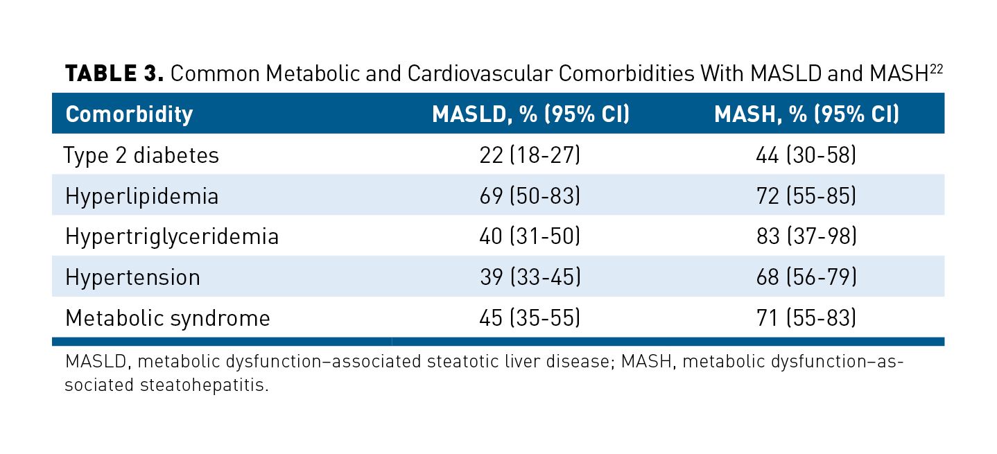
MASH is more common in patients with obesity and with T2D, but it can also occur in the absence of obesity and diabetes. Conversely, MASH is not present in all people with obesity and T2D. However, frequent comorbidities among people with MASLD and MASH include T2D, obesity, hyperlipidemia, hypertriglyceridemia, hypertension, and metabolic syndrome (Table 3).22 The most prevalent comorbidity in people with MASLD is hyperlipidemia (elevated cholesterol and/or triglyceride level).22 The most prevalent comorbidity among people with MASH is hypertriglyceridemia.22 Components of the metabolic syndrome generally were more prevalent among people with MASH than among those with MASLD.22
The presence of comorbidities including T2D, cardiovascular disease (CVD), and cirrhosis also increases the risk of death among patients with MASH.14 In an analysis of real-world outcomes among 28,576 patients with MASH in the US using Optum’s Clinfor-matics Data Mart Database (October 2015 to December 2022), the risk of all-cause death was nearly 5 times higher among patients with cirrhosis (HR, 4.68; 95% CI, 4.29-5.12) than among those without cirrhosis after adjustment for several baseline characteristics and comorbidities.14 The adjusted risk of all-cause death is increased with male sex (HR, 1.20; 95% CI, 1.12-1.29) and baseline T2D (HR, 1.78; 95% CI, 1.65-1.92) or CVD (HR, 1.58; 95% CI, 1.40-1.78).14 Among patients without cirrhosis at baseline, 23.1% experienced a composite clinical outcome of all-cause death or a significant hepatic event (including cirrhosis, decompensation, or liver transplant) over follow-up (mean: patients without cirrhosis, 3.2 years; patients with cirrhosis, 2.5 years), and the risk of progressing to this outcome increased with baseline T2D (HR, 1.28; 95% CI, 1.20-1.37), CVD (HR, 1.28; 95% CI, 1.19-1.38), or obesity (HR, 1.07; 95% CI, 1.00-1.15).14
The leading cause of mortality in patients with MASH is CVD, independent of other metabolic comorbidities.68 In an observational, longitudinal study conducted using National Health and Nutrition Examination Survey data (1988-1994) linked to National Death Index mortality outcomes (through 2019), the major drivers of mortality among patients with high-risk MASLD were CVD, cancer, and other-cause (likely liver-related) complications. Notably, the contribution of CVD increased with age.69
CV complications including atherosclerotic CVD (ASCVD) may result from visceral adiposity, atherogenic dyslipidemia (low HDL-C level; elevated levels of triglycerides and small-dense low density lipoprotein cholesterol [LDL-C]), and insulin resistance with or without hyperglycemia.69 Absolute reductions in LDL-C levels correlate with fewer major CV events, especially among patients with elevated aminotransferase levels.14
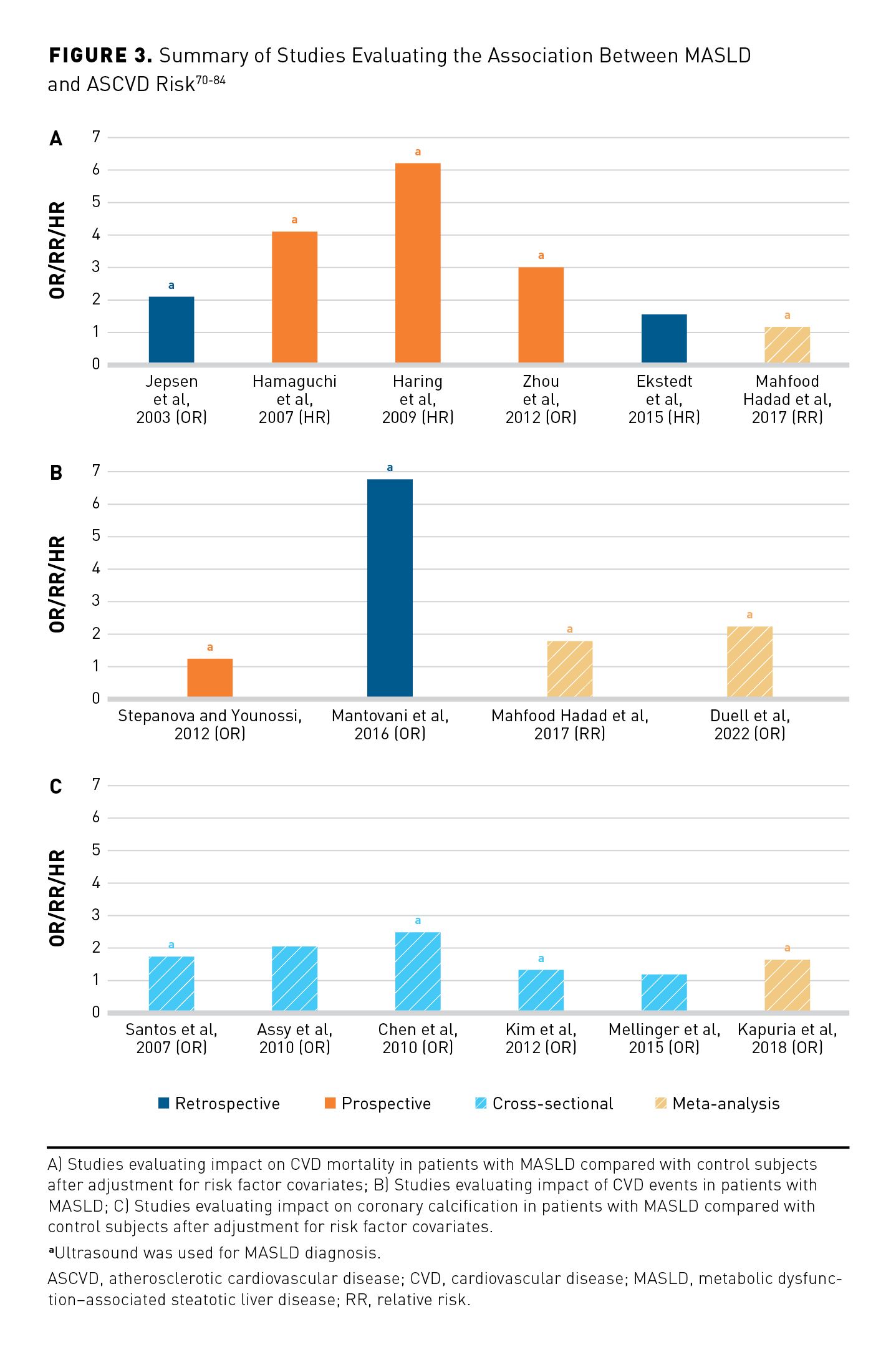
The American Heart Association has issued a scientific statement that includes a summary of study results involving the association between MASLD and ASCVD risk.70 The statement demonstrates that in several trials, MASLD is an underappreciated and independent risk factor for ASCVD even after adjustment for ASCVD risk factor covariates (Figure 3).70-84
Guideline- and Guidance-Based Approaches for Identification and Risk Stratification of MASH and Comorbidities
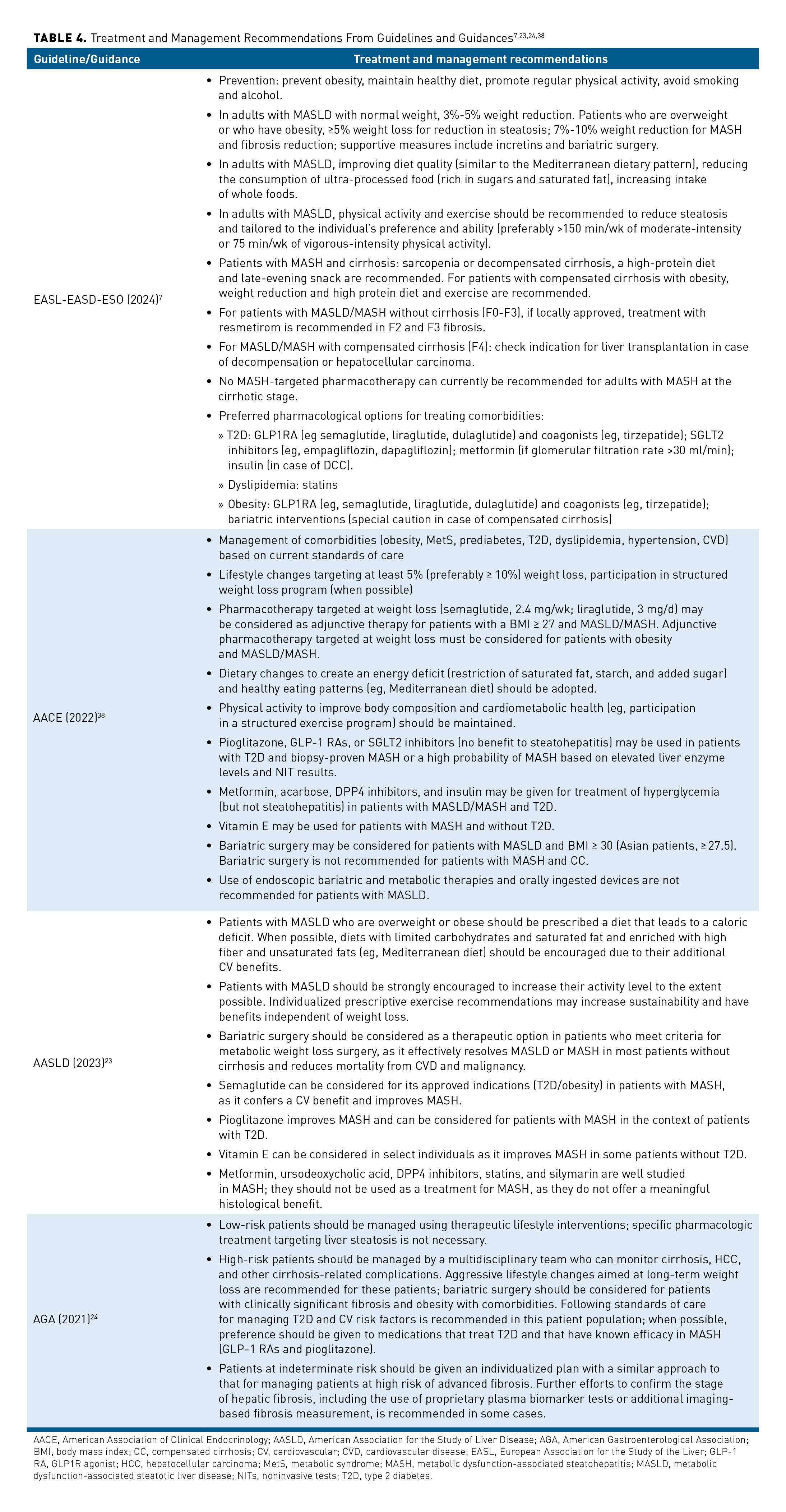
Guidelines and guidances on the overall management and risk stratification of MASLD and MASH were outlined most recently by the EASL/European Association for the Study of Diabetes/European Association for the Study of Obesity in 2024, as well as by the AASLD and AACE; these are summarized in Table 4.7,23,24,38 Additionally, the AGA has published guidance on the clinical care pathway for the risk stratification and management of patients with MASLD. The pathway stratifies patients by their risk of advanced fibrosis. Low risk of advanced fibrosis is defined as an FIB-4 score below 1.3, LSM below 8.0 kPa by transient elastography (TE), or a liver biopsy fibrosis stage of F0 to F1. Indeterminate risk is defined as an FIB-4 score of at least 1.3 and up to 2.67 and/or an LSM between 8.0 and 12.0 kPa on TE. High risk is defined as an FIB-4 score above 2.67, LSM above 12.0 kPa by TE, or a liver biopsy that shows clinically significant liver fibrosis (F2-F4).24 The AASLD guidance uses intermediate risk rather than indeterminate risk to denote FIB-4 scores of at least 1.3 and up to 2.67.23
New and Developmental Treatments
MASH is increasingly recognized as a major public health concern, reflecting the global rise in obesity and metabolic syndrome. Characterized by hepatic steatosis, inflammation, and varying degrees of fibrosis, MASH can progress to cirrhosis, hepatocellular carcinoma (HCC), and liver-related death. Significant fibrosis (F2-F4) in patients with MASH marks a critical threshold beyond which the risk of adverse outcomes escalates sharply. Therefore, timely identification and treatment of MASH with significant fibrosis are imperative to prevent irreversible liver damage and improve patient prognosis.
The current management of MASH focuses on more indirect causes of MASH and includes lifestyle interventions such as modifications to physical activity and diet that target weight loss and improved CV fitness.23,24 Before March 2024, in the absence of an approved MASH treatment, only the drugs used for comorbidities associated with MASH were included in guidance documents.23,24 However, the evidence for off-label use of such products in patients with MASH is limited, and administration of these agents does not provide benefits for all components of the disease (including no evidence of fibrosis improvement).23,24
Resmetirom (Resmetirom (MGL-3196; Madrigal Pharmaceuticals) is the first and only medicine approved by the FDA for the treatment of adult patients with NASH (MASH) accompanied by liver fibrosis.85 Resmetirom is a liver-directed, orally active, partial agonist for the THR with an approximately 28-fold selectivity for THR-β relative to THR-α compared to T3.85 Because resmetirom is a preferential THR-β agonist that has selective uptake into the liver, it avoids the systemic effects mediated by THR-α outside the liver.85 THR-β stimulation in the liver improves mitochondrial function and lipid metabolism and reduces blood LDL-C, apolipoprotein B, and triglyceride levels.85 Resmetirom was granted priority review, fast track, and breakthrough therapy designations by the FDA,86,87 and it was subsequently approved in conjunction with diet and exercise to treat adults with noncirrhotic nonalcoholic steatohepatitis (NASH/MASH) with moderate to advanced liver fibrosis (consistent with stages F2-F3 fibrosis).85
MAESTRO-NASH (NCT03900429) is an ongoing (as of publication) phase 3 trial of adults with biopsy-confirmed MASH and fibrosis stages F1B, F2, or F3.88 Patients were randomly assigned 1:1:1 to receive resmetirom, 80 or 100 mg, or placebo. The 2 primary end points at week 52 were NASH (MASH) resolution that included a reduction in the NAFLD activity score by at least 2 points with no worsening of fibrosis and a reduction in fibrosis by at least 1 stage with no worsening of the MASLD activity score. MASH resolution with no worsening of fibrosis was achieved in 25.9% of the patients in the group given 80 mg of resmetirom, 29.9% of those in the group given 100 mg of resmetirom, and 9.7% of those in the group given placebo (P < .001 for both comparisons with placebo). Fibrosis improvement by at least 1 stage with no worsening of the NAFLD activity score was achieved in 24.2%, 25.9, and 14.2% of patients, respectively (P < .001 for both comparisons with placebo). These data formed the basis of support for the accelerated approval of resmetirom for use in this patient population. The MAESTRONASH study is continuing through 2026.88
Treatments in Development
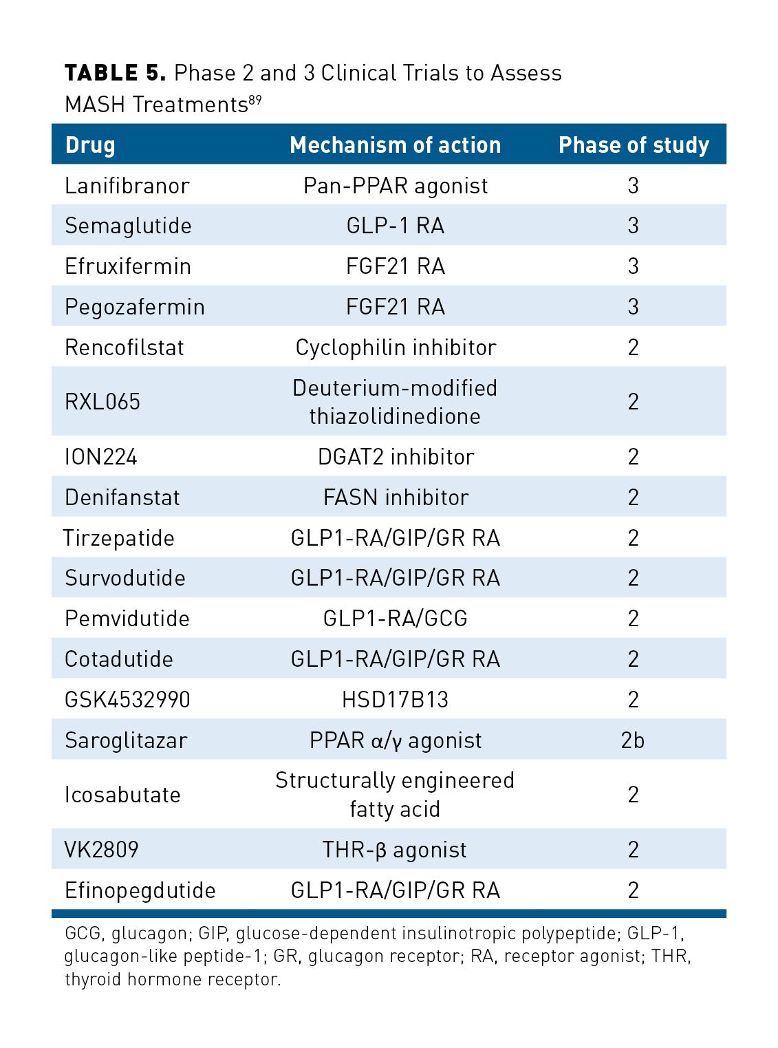
As of September 2023, multiple therapeutics with different mechanisms of action to treat MASH are being assessed in clinical trials (Table 5).89
Symptoms and Disease Burden
Liver damage has a deleterious impact on an individual’s quality of life (QOL) and general health status, with negative effects on QOL beginning early in the disease course. As MASH worsens, its symptoms— including anxiety, depression, fatigue, right upper quadrant pain, and impaired sleep, focus, and memory—often will hinder a patient’s ability to work and perform standard day-to-day tasks.90,91
As many as 80% of patients with MASH experience at least 1 other comorbidity. The most common comorbidities of MASH include obesity, CVD, and T2D, which lead to increased burden on patients and health systems.92
Economically, use of health care resources by patients with MASH has a widespread impact—the annual direct health care costs exceed $223 billion in the United States alone.92 With MASH prevalence increasing in the United States and globally, this economic burden is also expected to rise.92 On a per-patient basis, the direct costs of MASH (not including any comorbid, nonmedical, productivity, or societal costs) have been modeled at more than $32,000 over a patient’s lifetime, with costs increasing as the patient ages.92 There is also a correlation between the severity of disease as indicated by FIB-4 scores and the associated health care costs. In a claims-based, retrospective analysis of more than 6700 patients with MASH, there was a mean increase from $16,744 to $34,667 between the lowest and highest FIB-4 cohorts. A 1-unit increase in FIB-4 scores corresponds to a 3.4% increase in mean total annual costs, emphasizing the financial burden associated with MASH severity.13
Patients with MASH experience higher hospitalization rates than do patients in the general population and those with T2D alone.91 The combination of MASH with comorbidities amplifies health care costs compared with expenditures for MASH or comorbidities alone, with MASH and T2D leading to a substantial 63% incremental increase in expenses.30 With MASH likely to become the leading cause of advanced cirrhosis in coming decades, it is also predicted to become the leading cause of liver transplantation. Patients who undergo liver transplantation for MASH experience posttransplant hospitalization of up to 45 days. With an increasing number of liver transplantations attributable to MASH, the allocation of resources for this surgery will escalate.92
Conclusion
Screening and diagnosis of MASLD and MASH evolved from primarily biopsy-based methods to incorporate the use of noninvasive techniques. This progression has been reflected in several consensus evidence-based guidelines as more evidence to support the use of these techniques has been published.23,24 Some guidelines have provided additional detail on the use of biomarkers and other noninvasive approaches to stage disease severity, prognosis, and response to treatment.
Historically, the standard of care for managing MASH focused more on indirect causes of MASH and included lifestyle interventions such as modifications to physical activity and diet to promote weight loss.23,24 Until 2024, in the absence of a product approved by the FDA to treat NASH, pharmaceutical interventions to manage comorbidities also were recommended. However, the evidence for off-label use of such products in MASH is limited, and these agents do not provide benefits for all components of the disease (including no evidence of fibrosis improvement).23,24
Delayed treatment of MASH with significant fibrosis can lead to a cascade of adverse clinical outcomes. Patients with significant fibrosis are at a higher risk of developing cirrhosis, which is often irreversible and associated with complications such as variceal bleeding, ascites, hepatic encephalopathy, and HCC. Furthermore, cirrhotic patients face increased morbidity and mortality, reduced QOL, and a greater burden on health care resources. The natural history of MASH underscores the critical window of opportunity to intervene before advanced fibrosis and cirrhosis develop. Early and aggressive treatment strategies can significantly alter disease trajectory, emphasizing the need for heightened clinical awareness and proactive management.
The substantial use of health care resources, economic burden, and overall impact on patients underscore the need for a metabolism- and liver-directed treatment that may attenuate the progression of liver disease. Accordingly, the approval of resmetirom in March 2024 holds promise as a treatment for patients with MASH and MASLD and continues the evolution of screening, diagnosis, and management of the condition.
Disclosures of Financial Interest: Dr Noureddin reports: Advisory board for 89BIO, Altimmune, Boehringer Ingelheim, CytoDyn, GSK, Madrigal, Merck, Novo Nordisk, Terns, and Takeda. Principal investigator for a drug study for Akero, Allergan, Boehringer Ingelheim, Bristol Myers Squibb, Conatus, Corcept, Enanta, Galectin, GENFIT, Gilead, GSK, Madrigal, Novartis, Novo Nordisk, Shire, Takeda, Terns, Viking, and Zydus. Stockholder of ChronWell, CytoDyn, and Rivus Pharma.
Dr Alkhouri reports: Consultant for 89Bio, Boehringer Ingelheim, Echosens, Fibronostics, Gilead, Intercept, Ipsen, Madrigal, NorthSea, Novo Nordisk, Perspectum, Pfizer, and Zydus. Grant/research support from 89Bio, Akero, Arbutus, AstraZeneca, Better Therapeutics, Boehringer Ingelheim, Bristol Myers Squibb, Corcept, CymaBay, DSM, Galectin, Genentech, GENFIT, Gilead, Healio, Hepagene, Intercept, Inventiva, Ionis, Ipsen, Madrigal, Merck, NGM, Noom, NorthSea, Novo Nordisk, Perspectum, Pfizer, Poxel, Viking, and Zydus. Speaker’s fees from AbbVie, Alexion, Echosens, Gilead, Intercept, Ipsen, Madrigal, Perspectum, and Theratechnologies.
Funding: Support for this publication was provided by Madrigal Pharmaceuticals.
Acknowledgments: Editorial and writing support was provided by Thomas King, MPH, from Madrigal Pharmaceuticals.
Authorship Affiliation: Arizona Liver Health (NA), Phoenix, AZ; Houston Methodist Hospital and Houston Research Institute (MN), Houston, TX.
Source of Funding: This supplement was supported by Madrigal Pharmaceuticals.
Author Disclosures: Dr Alkhouri reports participation in consultancies or paid advisory boards with Echosens; Fibronostics; Gilead Sciences, Inc; Intercept Pharmaceuticals, Inc; Madrigal Pharmaceuticals; Novo Nordisk; Viking Therapeutics; and Zydus Pharmaceuticals, Inc. He also reports grants received from 89Bio, Inc; Boehringer Ingelheim; Gilead Sciences, Inc; Intercept Pharmaceuticals, Inc; Ionis Pharmaceuticals, Inc; Madrigal Pharmaceuticals; Novo Nordisk; Viking Therapeutics; and Zydus Pharmaceuticals, Inc. He has also received lecture fees for speaking at the invitation of Echosens; Gilead Sciences, Inc; and Intercept Pharmaceuticals, Inc. Dr Noureddin reports participating in consultancies or paid advisory boards for Aligos Therapeutics; Altimmune; AstraZeneca; Boehringer Ingelheim; Boston Pharmaceuticals; CytoDyn Inc; GSK plc; Lilly; Madrigal Pharmaceuticals; Merck & Co, Inc; Novo Nordisk; Takeda Pharmaceutical Company Limited; and Terns Pharmaceuticals, Inc. He reports lecture fees for speaking at the invitation of Madrigal Pharmaceuticals. He also reports stock ownership in ChronWell, CytoDyn Inc, and Rivus Pharmaceuticals. He reports being the principal investigator in a drug study with Akero Therapeutics, Inc; Allergan Pharmaceuticals; Boehringer Ingelheim; Bristol Myers Squibb; Conatus Pharma; Corcept Therapeutics, Inc; Enanta Pharmaceuticals, Inc; Galectin Therapeutics; Genfit; Gilead Sciences, Inc; GSK plc; Madrigal Pharmaceuticals; Novartis; Novo Nordisk; Takeda Pharmaceutical Company Limited; Terns Pharmaceuticals, Inc; Viking Therapeutics; and Zydus Pharmaceuticals, Inc.
Authorship Information: Concept and design (NA, MN); drafting of the manuscript (NA, MN); critical revision of the manuscript for important intellectual content (NA, MN).
Address Correspondence To: Naim Alkhouri, MD, FAASLD. Arizona Liver Health, Tucson Liver Clinic, 1601 North Swan Road, Tucson, AZ 85712. Email: naim.alkhouri@gmail.com
REFERENCES
1. Ludwig J, Viggiano TR, McGill DB, Oh BJ. Nonalcoholic steatohepatitis: Mayo Clinic experiences with a hitherto unnamed disease. Mayo Clin Proc. 1980;55(7):434-438.
2. Kanwall F, Neuschwander-Tetri BA, Loomba R, Rinella ME. Metabolic dysfunction-associated steatotic liver disease (MASLD): update and impact of new nomenclature on the AASLD clinical practice guidance on nonalcoholic fatty liver disease. Hepatology. 2024;79(5):1212-1219. doi:10.1097/HEP.0000000000000670
3. Song SJ, Lai JC, Wong GL, Wong VW, Yip TC. Can we use old NAFLD data under the new MASLD definition? J Hepatol. 2024;80(2):e54-e56 doi:10.1016/j.jhep.2023.07.021
4. Ratziu V, Boursier J; AFEF Group for the Study of Liver Fibrosis. Confirmatory biomarker diagnostic studies are not needed when transitioning from NAFLD to MASLD. J Hepatol. 2024;80(2):e51-e52. doi:10.1016/j.jhep.2023.07.017
5. Chalasani N, Younossi Z, Lavine JE, et al. The diagnosis and management of nonalcoholic fatty liver disease: practice guidance from the American Association for the Study of Liver Diseases. Hepatology. 2018;67(1):328-357. doi:10.1002/hep.29367
6. Powell EE, Wong VW, Rinella M. Non-alcoholic fatty liver disease. Lancet. 2021;397(10290):2212-2224. doi:10.1016/s0140-6736(20)32511-3
7. European Association for the Study of the Liver (EASL); European Association for the Study of Diabetes (EASD); European Association for the Study of Obesity (EASO). EASL-EASD-EASO clinical practice guidelines on the management of metabolic dysfunction-associated steatotic liver disease (MASLD). J Hepatol. Published online June 5, 2024. doi:10.1016/j.jhep.2024.04.031
8. Review Team; LaBrecque DR, Abbas Z, et al; World Gastroenterology Organisation. World Gastroenterology Organisation global guidelines: nonalcoholic fatty liver disease and nonalcoholic steatohepatitis. J Clin Gastroenterol. 2014;48(6):467-473. doi:10.1097/mcg.0000000000000116
9. Monelli F, Venturelli F, Bonilauri L, et al. Systematic review of existing guidelines for NAFLD assessment. Hepatoma Res. 2021;7:25. doi:10.20517/2394-5079.2021.03
10. Tokushige K, Ikejima K, Ono M, et al. Evidence-based clinical practice guidelines for nonalcoholic fatty liver disease/nonalcoholic steatohepatitis 2020. J Gastroenterol. 2021;56(11):951-963. doi:10.1007/s00535-021-01796-x
11. Associazione Italiana per lo Studio del Fegato (AISF), Società Italiana di Diabetologia (SID), and Società Italiana dell’Obesità (SIO); Members of the guidelines panel; Coordinator; et al. Non-alcoholic fatty liver disease in adults 2021: a clinical practice guideline of the Italian Association for the Study of the Liver (AISF), the Italian Society of Diabetology (SID), and the Italian Society of Obesity (SIO). Dig Liver Dis. 2021; 54(2): 170-182. doi:10.1016/j.dld.2021.04.029
12. Nonalcoholic fatty liver disease (NAFLD) & NASH. National Institute of Diabetes and Digestive and Kidney Diseases. Reviewed April 2021. Accessed August 14, 2024. https://www.niddk.nih.gov/healthinformation/liver-disease/nafld-nash
13. Tapper EB, Bonafede M, Fishman J, et al. Healthcare resource utilization and costs of care in the United States for patients with non-alcoholic steatohepatitis. J Med Econ. 2023;26(1):348-356. doi:10.1080/13696998.2023.2184967
14. Charlton M, Qian C, Szabo S, et al. Characterizing the management of patients with NASH (with versus without cirrhosis) in real-world clinical practice—low utilization of gastroenterology and hepatology specialty care. Abstract presented at the Therapeutic Agents for Non-Alcoholic Steatohepatitis (NASHTAG) 2024 Meeting; January 4-6, 2024; Park City, Utah. Abstract 25.
15. Harrison SA, Ratziu V, Anstee QM, et al. Design of the phase 3 MAESTRO clinical program to evaluate resmetirom for the treatment of nonalcoholic steatohepatitis. Aliment Pharmacol Ther. 2024;59(1):51-63. doi:10.1111/apt.17734
16. Anstee QM, Hallsworth K, Lynch N, et al. Real-world management of non-alcoholic steatohepatitis differs from clinical practice guideline recommendations and across regions. JHEP Rep. 2022;4(1):100411. doi:10.1016/j.jhepr.2021.100411
17. Loomba R, Ratziu V, Harrison SA; NASH Clinical Trial Design International Working Group. Expert panel review to compare FDA and EMA guidance on drug development and endpoints in nonalcoholic steatohepatitis. Gastroenterology. 2022;162(3):680-688. doi:10.1053/j.gastro.2021.10.051
18. Ratziu V, Charlotte F, Heurtier A, et al. Sampling variability of liver biopsy in nonalcoholic fatty liver disease. Gastroenterology. 2005;128(7):1898-1906. doi:10.1053/j.gastro.2005.03.084
19. Seeff LB, Everson GT, Morgan TR, et al; HALT-C Trial Group. Complication rate of percutaneous liver biopsies among persons with advanced chronic liver disease in the HALT-C trial. Clin Gastroenterol Hepatol. 2010;8(10):877-883. doi:10.1016/j.cgh.2010.03.025
20. Chowdhury AB, Mehta KJ. Liver biopsy for assessment of chronic liver diseases: a synopsis. Clin Exp Med. 2023;23(2):273-285. doi:10.1007/s10238-022-00799-z
21. Grandison GA, Angulo P. Can NASH be diagnosed, graded, and staged noninvasively? Clin Liver Dis. 2012;16(3):567-585. doi:10.1016/j.cld.2012.05.001
22. Younossi ZM, Koenig AB, Abdelatif D, Fazel Y, Henry L, Wymer M. Global epidemiology of nonalcoholic fatty liver disease-Meta-analytic assessment of prevalence, incidence, and outcomes. Hepatology. 2016;64(1):73-84. doi:10.1002/hep.28431
23. Rinella ME, Neuschwander-Tetri BA, Siddiqui MS, et al. AASLD practice guidance on the clinical assessment and management of nonalcoholic fatty liver disease. Hepatology. 2023;77(5):1797-1835. doi:10.1097/HEP.0000000000000323
24. Kanwal F, Shubrook JH, Adams LA, et al. Clinical care pathway for the risk stratification and management of patients with nonalcoholic fatty liver disease. Gastroenterology. 2021;161(5):1657-1669. doi:10.1053/j.gastro.2021.07.049
25. Harrison SA, Gawrieh S, Roberts K, et al. Prospective evaluation of the prevalence of non-alcoholic fatty liver disease and steatohepatitis in a large middle-aged US cohort. J Hepatol. 2021;75(2):284-291. doi:10.1016/j.jhep.2021.02.034
26. Younossi ZM, Blissett D, Blissett R, et al. The economic and clinical burden of nonalcoholic fatty liver disease in the United States and Europe. Hepatology. 2016;64(5):1577-1586. doi:10.1002/hep.28785
27. Diehl AM, Day C. Cause, pathogenesis, and treatment of nonalcoholic steatohepatitis. N Engl J Med. 2017;377(21):2063-2072. doi:10.1056/NEJMra1503519
28. Rich NE, Oji S, Mufti AR, et al. Racial and ethnic disparities in nonalcoholic fatty liver disease prevalence, severity, and outcomes in the United States: a systematic review and meta-analysis. Clin Gastroenterol Hepatol. 2018;16(2):198-210.e2. doi:10.1016/j.cgh.2017.09.041
29. Census bureau projects U.S. and world populations on New Year’s Day. News release. United States Census Bureau. December 29, 2022. Accessed August 14, 2024. https://www.census.gov/newsroom/press-releases/2022/new-years-day-population.html
30. Fishman J, Tapper EB, Dodge S, et al. The incremental cost of non-alcoholic steatohepatitis and type 2 diabetes in the United States using real-world data. Curr Med Res Opin. 2023;39(11):145-1429. doi:10.1080/03007995.2023.2262926
31. Ahmed O, Liu L, Gayed A, et al. The changing face of hepatocellular carcinoma: forecasting prevalence of nonalcoholic steatohepatitis and hepatitis C cirrhosis. J Clin Exp Hepatol. 2019;9(1):50-55. doi:10.1016/j.jceh.2018.02.006
32. Estes C, Anstee QM, Arias-Loste MT, et al. Modeling NAFLD disease burden in China, France, Germany, Italy, Japan, Spain, United Kingdom, and United States for the period 2016-2030. J Hepatol. 2018;69(4):896-904. doi:10.1016/j.jhep.2018.05.036
33. Kabbany MN, Conjeevaram Selvakumar PK, Watt K, et al. Prevalence of nonalcoholic steatohepatitis-associated cirrhosis in the United States: an analysis of National Health and Nutrition Examination Survey data. Am J Gastroenterol. 2017;112(4):581-587. doi:10.1038/ajg.2017.5
34. Buzzetti E, Pinzani M, Tsochatzis EA. The multiple-hit pathogenesis of non-alcoholic fatty liver disease (NAFLD). Metabolism. 2016;65(8):1038-1048. doi:10.1016/j.metabol.2015.12.012
35. Mota M, Banini BA, Cazanave SC, Sanyal AJ. Molecular mechanisms of lipotoxicity and glucotoxicity in nonalcoholic fatty liver disease. Metabolism. 2016;65(8):1049-1061. doi:10.1016/j.metabol.2016.02.014
36. Chaves C, Bruinstroop E, Refetoff S, Yen PM, Anselmo J. Increased hepatic fat content in patients with resistance to thyroid hormone beta. Thyroid. 2021;31(7):1127-1134. doi:10.1089/thy.2020.0651
37. Zarei M, Aguilar-Recarte D, Palomer X, Vázquez-Carrera M. Revealing the role of peroxisome proliferator-activated receptor β/δ in nonalcoholic fatty liver disease. Metabolism. 2021;114:154342. doi:10.1016/j.metabol.2020.154342
38. Cusi K, Isaacs S, Barb D, et al. American Association of Clinical Endocrinology clinical practice guideline for the diagnosis and management of nonalcoholic fatty liver disease in primary care and endocrinology clinical settings: co-sponsored by the American Association for the Study of Liver Diseases (AASLD). Endocr Pract. 2022;28(5):528-562. doi:10.1016/j.eprac.2022.03.010
39. Davison BA, Harrison SA, Cotter G, et al. Suboptimal reliability of liver biopsy evaluation has implications for randomized clinical trials. J Hepatol. 2020;73(6):1322. doi:10.1016/j.jhep.2020.06.025
40. McPherson S, Hardy T, Dufour JF, et al. Age as a confounding factor for the accurate non-invasive diagnosis of advanced NAFLD fibrosis. Am J Gastroenterol. 2017;112(5):740-751. doi:10.1038/ajg.2016.453
41. Bernstein D, Kovalic AJ. Noninvasive assessment of fibrosis among patients with nonalcoholic fatty liver disease [NAFLD]. Metabol Open. 2022;13:100158. doi:10.1016/j.metop.2021.100158
42. Jung J, Loomba RR, Imajo K, et al. MRE combined with FIB-4 (MEFIB) index in detection of candidates for pharmacological treatment of NASH-related fibrosis. Gut. 2021;70(10):1946-1953. doi:10.1136/gutjnl-2020-322976
43. Noureddin M, Truong E, Gornbein JA, et al. MRI-based (MAST) score accurately identifies patients with NASH and significant fibrosis. J Hepatol. 2022;76(4):781-787. doi:10.1016/j.jhep.2021.11.012
44. Gidener T, Ahmed OT, Larson JJ, et al. Liver stiffness by magnetic resonance elastography predicts future cirrhosis, decompensation, and death in NAFLD. Clin Gastroenterol Hepatol. 2021;19(9):1915-1924. e6. doi:10.1016/j.cgh.2020.09.044
45. Dulai PS, Sirlin CB, Loomba R. MRI and MRE for non-invasive quantitative assessment of hepatic steatosis and fibrosis in NAFLD and NASH: clinical trials to clinical practice. J Hepatol. 2016;65(5):1006-1016. doi:10.1016/j.jhep.2016.06.005
46. Beyer C, Hutton C, Andersson A, et al. Comparison between magnetic resonance and ultrasound-derived indicators of hepatic steatosis in a pooled NAFLD cohort. PLoS One. 2021;16(4):e0249491. doi:10.1371/journal.pone.0249491
47. Tamaki N, Munaganuru N, Jung J, et al. Clinical utility of 30% relative decline in MRI-PDFF in predicting fibrosis regression in non-alcoholic fatty liver disease. Gut. 2022;71(5):983-990. doi:10.1136/gutjnl-2021-324264
48. Fujii H, Fukumoto S, Enomoto M, et al. The FibroScan-aspartate aminotransferase score can stratify the disease severity in a Japanese cohort with fatty liver diseases. Sci Rep. 2021;11(1):13844. doi:10.1038/s41598-021-93435-x
49. Newsome PN, Sasso M, Deeks JJ, et al. FibroScan-AST (FAST) score for the non-invasive identification of patients with non-alcoholic steatohepatitis with significant activity and fibrosis: a prospective derivation and global validation. Lancet Gastroenterol Hepatol. 2020;5(4):362-373. doi:10.1016/S2468-1253(19)30383-8
50. SpeakLiver: Understanding MASH. Accessed November 1, 2024. https://www.speakliver.com/what-ismash-liver-disease.html
51. Sharma B, John S. Nonalcoholic steatohepatitis (NASH). StatPearls. National Center for Biotechnology Information. Updated April 7, 2023. Accessed August 14, 2024. https://www.ncbi.nlm.nih.gov/books/NBK470243/
52. Stengel JZ, Harrison SA. Nonalcoholic steatohepatitis: clinical presentation, diagnosis, and treatment. Gastroenterol Hepatol (N Y). 2006;2(6):440-449.
53. Nagra N, Penna R, La Selva D, Coy D, Siddique A, Burman B. Tagging incidental finding of fatty liver on ultrasound: a novel intervention to improve early detection of liver fibrosis. J Clin Transl Res. 2021;7(5):641-647.
54. Yang X, Chang X, Wu S, et al. Performance of liver stiffness measurements obtained with FibroScan is affected by glucose metabolism in patients with nonalcoholic fatty liver disease. Lipids Health Dis. 2021;20(1):27. doi:10.1186/s12944-021-01453-5
55. Xie Q, Zhou X, Huang P, Wei J, Wang W, Zheng S. The performance of enhanced liver fibrosis (ELF) test for the staging of liver fibrosis: a meta-analysis. PLoS One. 2014;9(4):e92772. doi:10.1371/journal.pone.0092772
56. Stine JG, Munaganuru N, Barnard A, et al. Change in MRI-PDFF and histologic response in patients with nonalcoholic steatohepatitis: a systematic review and meta-analysis. Clin Gastroenterol Hepatol. 2021;19(11):2274-2283.e5. doi:10.1016/j.cgh.2020.08.061
57. Newsome PN, Cramb R, Davison SM, et al. Guidelines on the management of abnormal liver blood tests. Gut. 2018;67(1):6-19. doi:10.1136/gutjnl-2017-314924
58. FDA ‘breakthrough device’ designation for diagnosis granted for Siemens Healthineers enhanced liver fibrosis test, signifies progress for nonalcoholic fatty liver disease. News release. Siemens Healthineers. July 18, 2023. Accessed August 14, 2024. https://siemens-healthineers.com/en-us/press-room/pressreleases/elf-test-breakthrough-device-designation-diagnosis
59. Mak AL, Lee J, van Dijk AM, et al. Systematic review with meta-analysis: diagnostic accuracy of Pro- C3 for hepatic fibrosis in patients with non-alcoholic fatty liver disease. Biomedicines. 2021;9(12):1920. doi:10.3390/biomedicines9121920
60. Harrison SA, Ratziu V, Magnanensi J, et al. NIS2+, an optimisation of the blood-based biomarker NIS4 technology for the detection of at-risk NASH: a prospective derivation and validation study. J Hepatol. 2023;79(3):758-767. doi:10.1016/j.jhep.2023.04.031
61. Boursier J, Hagström H, Ekstedt M, et al. Non-invasive tests accurately stratify patients with NAFLD based on their risk of liver-related events. J Hepatol. 2022;76(5):1013-1020. doi:10.1016/j.jhep.2021.12.031
62. Zhang YN, Fowler KJ, Hamilton G, et al. Liver fat imaging-a clinical overview of ultrasound, CT, and MR imaging. Br J Radiol. 2018;91(1089):20170959. doi:10.1259/bjr.20170959
63. Chartampilas E. Imaging of nonalcoholic fatty liver disease and its clinical utility. Hormones (Athens). 2018;17(1):69-81. doi:10.1007/s42000-018-0012-x
64. Home page. Echosens. Accessed August 14, 2024. https://www.echosens.com/en-us
65. Mózes FE, Lee JA, Vali Y, et al; LITMUS Investigators. Performance of non-invasive tests and histology for the prediction of clinical outcomes in patients with non-alcoholic fatty liver disease: an individual participant data meta-analysis. Lancet Gastroenterol Hepatol. 2023;8(8):704-713. doi:10.1016/S2468-1253(23)00141-3
66. Selvaraj EA, Mózes FE, Jayaswal ANA, et al; LITMUS Investigators. Diagnostic accuracy of elastography and magnetic resonance imaging in patients with NAFLD: a systematic review and meta-analysis. J Hepatol. 2021;75(4):770-785. doi:10.1016/j.jhep.2021.04.044
67. Younossi ZM, Loomba R, Anstee QM, et al. Diagnostic modalities for nonalcoholic fatty liver disease, nonalcoholic steatohepatitis, and associated fibrosis. Hepatology. 2018;68(1):349-360. doi:10.1002/hep.29721
68. Shroff H, VanWagner LB. Cardiovascular disease in nonalcoholic steatohepatitis: screening and management. Curr Hepatol Rep. 2020;19(3):315-326. doi:10.1007/s11901-020-00530-0
69. Fishman J, Kim Y, Woolley J, Parrinello C, O’Connell T. Association of all-cause and cause-specific mortality with high-risk NAFLD/NASH in US adults, NHANES III (1988-1994) linked to mortality data through 2019. Value Health. 2024;27(6; suppl):S44. doi:10.1016/j.jval.2024.03.231
70. Duell PB, Welty FK, Miller M, et al; American Heart Association Council on Arteriosclerosis, Thrombosis and Vascular Biology; Council on Hypertension; Council on the Kidney in Cardiovascular Disease; Council on Lifestyle and Cardiometabolic Health; and Council on Peripheral Vascular Disease. Nonalcoholic fatty liver disease and cardiovascular risk: a scientific statement from the American Heart Association. Arterioscler Thromb Vasc Biol. 2022;42(6):e168-e185. doi:10.1161/ATV.0000000000000153
71. Jepsen P, Vilstrup H, Mellemkjaer L, et al. Prognosis of patients with a diagnosis of fatty liver—a registry-based cohort study. Hepatogastroenterology. 2003;50(54):2101-2104.
72. Hamaguchi M, Kojima T, Takeda N, et al. Nonalcoholic fatty liver disease is a novel predictor of cardiovascular disease. World J Gastroenterol. 2007;13(10):1579-1584. doi:10.3748/wjg.v13.i10.1579
73. Haring R, Wallaschofski H, Nauck M, Dörr M, Baumeister SE, Völzke H. Ultrasonographic hepatic steatosis increases prediction of mortality risk from elevated serum gamma-glutamyl transpeptidase levels. Hepatology. 2009;50(5):1403-1411. doi:10.1002/hep.23135
74. Zhou YJ, Li YY, Nie YQ, Huang CM, Cao CY. Natural course of nonalcoholic fatty liver disease in southern China: a prospective cohort study. J Dig Dis. 2012;13:153-160. doi:10.1111/j.1751-2980.2011.00571.x
75. Ekstedt M, Hagström H, Nasr P, et al. Fibrosis stage is the strongest predictor for disease-specific mortality in NAFLD after up to 33 years of follow-up. Hepatology. 2015;61:1547-1554. doi:10.1002/hep.27368
76. Mahfood Haddad T, Hamdeh S, Kanmanthareddy A, Alla VM. Nonalcoholic fatty liver disease and the risk of clinical cardiovascular events: a systematic review and meta-analysis. Diabetes Metab Syndr. 2017;11(suppl 1):S209-S216. doi:10.1016/j.dsx.2016.12.033
77. Stepanova M, Younossi ZM. Independent association between nonalcoholic fatty liver disease and cardiovascular disease in the US population. Clin Gastroenterol Hepatol. 2012;10:646-650. doi:10.1016/j.cgh.2011.12.039
78. Mantovani A, Mingolla L, Rigolon R, et al. Nonalcoholic fatty liver disease is independently associated with an increased incidence of cardiovascular disease in adult patients with type 1 diabetes. Int J Cardiol. 2016;225:387-391. doi:10.1016/j.ijcard.2016.10.040
79. Santos RD, Nasir K, Conceição RD, Sarwar A, Carvalho JA, Blumenthal RS. Hepatic steatosis is associated with a greater prevalence of coronary artery calcification in asymptomatic men. Atherosclerosis. 2007;194:517-519. doi:10.1016/j.atherosclerosis.2007.01.026
80. Assy N, Djibre A, Farah R, Grosovski M, Marmor A. Presence of coronary plaques in patients with nonalcoholic fatty liver disease. Radiology. 2010;254:393-400. doi:10.1148/radiol.09090769
81. Chen CH, Nien CK, Yang CC, Yeh YH. Association between nonalcoholic fatty liver disease and coronary artery calcification. Dig Dis Sci. 2010;55:1752-1760. doi:10.1007/s10620-009-0935-9
82. Kim D, Choi SY, Park EH, et al. Nonalcoholic fatty liver disease is associated with coronary artery calcification. Hepatology. 2012;56:605-613. doi:10.1002/hep.25593
83. Mellinger JL, Pencina KM, Massaro JM, et al. Hepatic steatosis and cardiovascular disease outcomes: an analysis of the Framingham Heart Study. J Hepatol. 2015;63:470-476. doi:10.1016/j.jhep.2015.02.045
84. Kapuria D, Takyar VK, Etzion O, Surana P, O’Keefe JH, Koh C. Association of hepatic steatosis with subclinical atherosclerosis: systematic review and meta-analysis. Hepatol Commun. 2018;2:873-883. doi:10.1002/hep4.1199
85. Rezdiffra. Prescribing information. Madrigal Pharmaceuticals, Inc; 2024. Accessed August 14, 2024. https://www.madrigalpharma.com/wp-content/uploads/2024/06/NDA-217785_REZDIFFRA-PI_14Mar2024_final-revised-clean-SPLPPI.pdf
86. Madrigal receives breakthrough therapy designation from FDA for resmetirom and completes enrollment of the phase 3 MAESTRO-NASH biopsy trial. News release. Madrigal Pharmaceuticals. April 18, 2024. Accessed August 4, 2024. https://ir.madrigalpharma.com/news-releases/news-release-details/madrigal-receives-breakthrough-therapy-designation-fda
87. Madrigal Pharmaceuticals announces NDA acceptance and priority review of the new drug application for resmetirom for the treatment of NASH with liver fibrosis. News release. Madrigal Pharmaceuticals. September 13, 2023. Accessed August 14, 2024. https://ir.madrigalpharma.com/news-releases/newsrelease-details/madrigal-pharmaceuticals-announces-nda-acceptance-and-priority
88. Harrison SA, Bedossa P, Guy CD, et al; MAESTRO-NASH Investigators. A phase 3, randomized, controlled trial of resmetirom in NASH with liver fibrosis. N Engl J Med. 2024;390(6):497-509. doi:10.1056/NEJMoa2309000
89. Harrison SA, Loomba R, Dubourg J, Ratziu V, Noureddin M. Clinical trial landscape in NASH. Clin Gastroenterol Hepatol. 2023;21(8):2001-2014. doi:10.1016/j.cgh.2023.03.041
90. Younossi Z, Aggarwal P, Shrestha I, et al. The burden of non-alcoholic steatohepatitis: a systematic review of health-related quality of life and patient-reported outcomes. JHEP Rep. 2022;4(9):100525. doi:10.1016/j.jhepr.2022.100525
91. Povsic M, Wong OY, Perry R, Bottomley J. A structured literature review of the epidemiology and disease burden of non-alcoholic steatohepatitis (NASH). Adv Ther. 2019;36(7):1574-1594. doi:10.1007/s12325-019-00960-3
92. Witkowski M, Moreno SI, Fernandes J, Johansen P, Augusto M, Nair S. The economic burden of nonalcoholic steatohepatitis: a systematic review. Pharmacoeconomics. 2022;40(8):751-776. doi:10.1007/s40273-022-01140-y
