- Center on Health Equity & Access
- Clinical
- Health Care Cost
- Health Care Delivery
- Insurance
- Policy
- Technology
- Value-Based Care
Evolving Knowledge of the Unmet Needs in Dry Eye Disease
Abstract
Dry eye disease is a common but underdiagnosed disorder in the United States, and its prevalence is likely to increase as the nation’s population ages. Although still regarded as little more than a nuisance by many clinicians and payers alike, dry eye disease is known to have both clinical consequences for ocular health and effects on vision-related quality of life in many people, impairing their ability to function well in vocational and social settings. Pharmaceutical treatments consist mainly of over-the-counter ocular lubricants (“artificial tears”) and a few prescription drugs that address the inflammatory component of dry eye disease through immunomodulation and/or inhibition of T-cell activity. In September 2020, Oyster Point Pharma, Inc (Oyster Point Pharma), convened a panel—consisting of 7 managed care executives with experience in management of dry eye disease treatments and 2 eye care practitioners with expertise in dry eye disease—to discuss how the growth of knowledge about dry eye disease in the past 2 decades has altered their thinking about and approach to dry eye disease, as well as how they would like to see the field advance. They pointed to an existing unmet need in knowledge and therapeutics that can address the underlying causes of dry eye disease. Oyster Point Pharma supported the authorship of this article; the authors were members of the panel, and all panelists were compensated by Oyster Point Pharma. This article provides an overview of dry eye disease and summarizes the panel discussion.
____________________________________________________________________________________________
Am J Manag Care. 2021;27(suppl 2):S23-S32. https://doi.org/10.37765/ajmc.2021.88625
For author information and disclosures, see end of text.
Introduction
Dry eye disease is a common, underdiagnosed disorder in the United States, and its prevalence is likely to increase as the nation’s population ages.1,2 It already is among the most frequent reasons people seek eye care.3 Dry eye disease adversely affects people’s health- and vision-related quality of life (QOL), impairing their ability to fully engage in vocational and leisurely activities and to perform normal activities of daily living.3 Because it affects millions of people in the workforce, dry eye disease may impose a substantial economic burden on the US health care system and society, largely in the form of reduced productivity.4 Compared with people without dry eye disease, people with clinically significant dry eye disease are about 2 to 3.5 times more likely to report problems with doing professional work, reading, using a computer, watching television, or driving.3 For these reasons, dry eye disease warrants attention from the various stakeholders involved in population health management.
In September 2020, Oyster Point Pharma, Inc (Oyster Point Pharma), assembled a panel to participate in a moderated discussion regarding advances in knowledge about dry eye disease and unmet needs in this field. The panel consisted of 7 managed care organization executives and 2 eye care practitioners with expertise in dry eye disease (an ophthalmologist who is a cornea cataract refractive surgeon and an optometrist who runs a large dry eye disease clinic where nearly all patients are seen by referral). Panelists were compensated by Oyster Point Pharma for their participation. A summary of their discussion follows the overview below covering dry eye disease disease state, epidemiology, and pathophysiology. Based in Princeton, New Jersey, Oyster Point Pharma is a clinical-stage biopharmaceutical company focused on the discovery, development, and commercialization of first-in-class pharmaceutical therapies to treat ocular surface diseases.
Evolving Definitions and Understanding of Dry Eye Disease
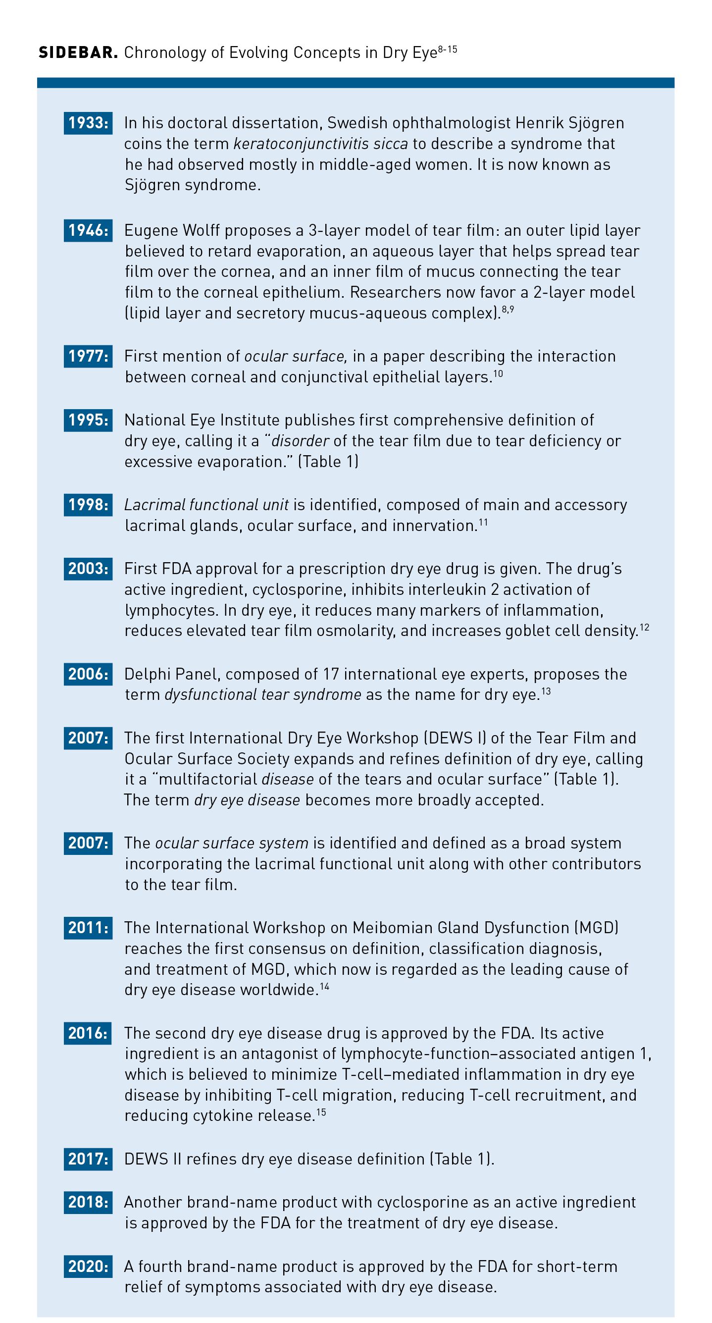
The concept of dry eye disease as a disease entity is relatively new. Definitions and concepts of dry eye disease have evolved over the past 25 years (Table 15-7; Sidebar8-15), reflecting the advances in knowledge that have emerged with a virtual explosion of scholarly publications about dry eye disease during this period.16
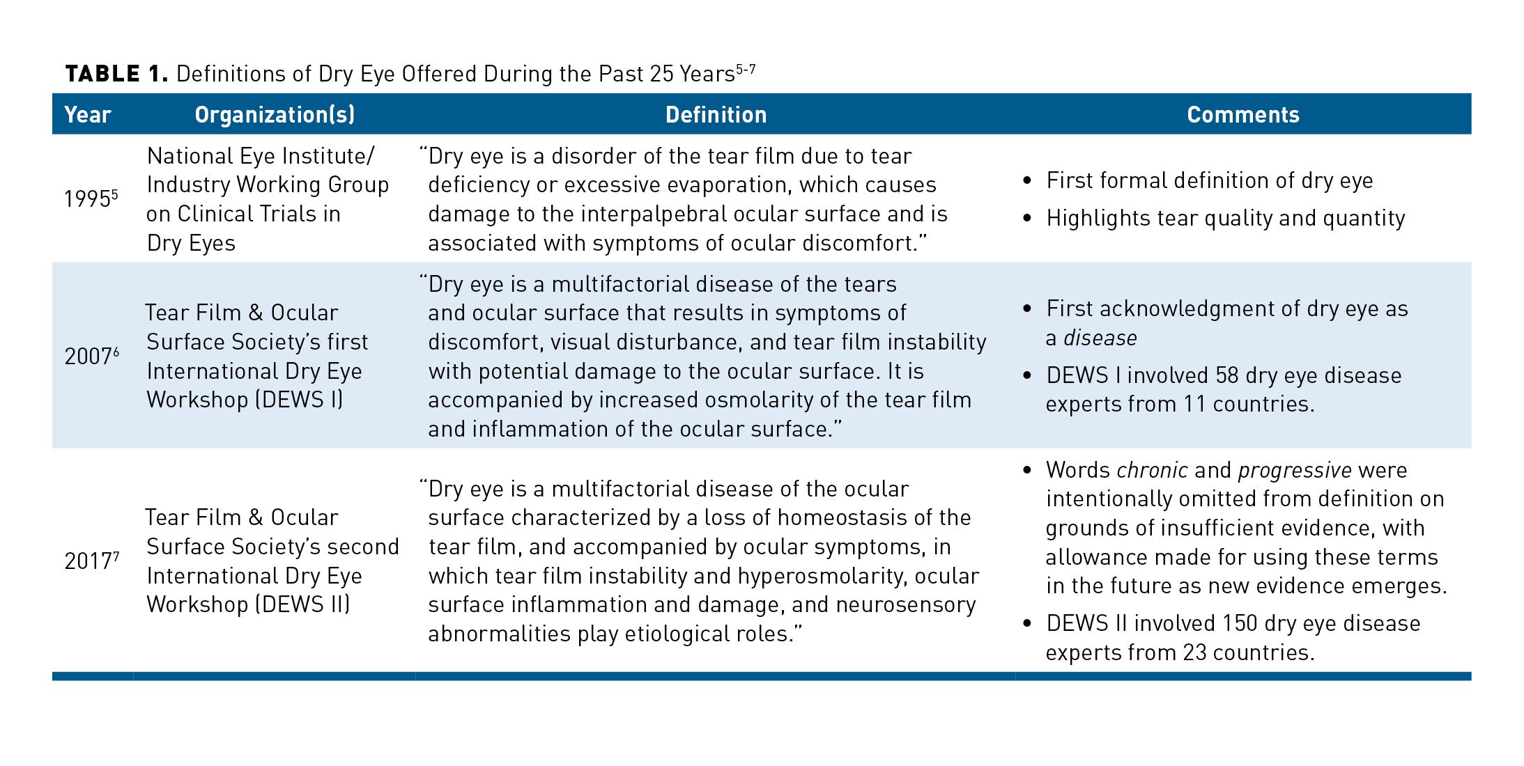
In its 1995 report, the National Eye Institute/Industry Workshop divided dry eye into 2 categories: tear-deficient dry eye, addressing tear quantity, and evaporative dry eye (EDE), addressing tear quality.5 Within tear-deficient dry eye were 2 subcategories: Sjögren syndrome dry eye and non-Sjögren tear-deficient dry eye. The latter condition stems from lacrimal disease, lacrimal obstruction, or reflex block. EDE was further classified according to etiologies involving oil deficiency, eyelid abnormalities, contact lens usage, and ocular surface change.
In 2007, the Tear Film & Ocular Surface Society sponsored the first International Dry Eye Workshop (DEWS I). The subsequent report mostly confirmed the above classification scheme but replaced tear-deficient with aqueous-deficient. These schemes led to the widespread misperception that the aqueous-deficient dry eye (ADDE) and EDE subtypes were mutually exclusive. In its 2017 report, DEWS II argued that instead of being either aqueous-deficient or evaporative, dry eye disease actually exists along a continuum, such that clinicians need to consider aspects of each subtype in the diagnosis and treatment of dry eye disease because a patient’s tears may be deficient in both quantity and quality.17
Epidemiology. In the absence of a standardized definition and classification system for dry eye disease, along with substantial differences in demographic characteristics among populations studied, the efforts to estimate the prevalence of dry eye disease according to symptoms, signs, or both have understandably produced widely varying results.18 A symptom-based US study, the Beaver Dam Offspring Study, found an overall prevalence of dry eye symptoms of 14.5%.19 Extrapolated without statistical adjustment to the 2019 US population 18 years and older (about 255 million people), this prevalence rate suggests that about 37 million adults in the United States may experience symptoms of dry eye disease.
In the Beaver Dam Offspring Study, potential participants were identified by their responses to these questions: “How often do you have dry eyes, a dry, gritty, or burning feeling?” “Is there a season of the year when the dryness in your eyes is the worst?” and “Are you currently using eyedrops at least once a day for dry eyes?” Participants who affirmatively answered the question about eyedrop usage and/or those who reported symptoms that were at least “moderately bothersome” and occurred “sometimes or more often” were identified as cases.
Results of another US study based on data from the 2013 National Health and Wellness Survey suggested that up to 9.3% of the adult population may experience dry eye disease, including the 6.8% of adult men and women (16.4 million) who received a diagnosis of dry eye disease.20 In this study, diagnosed dry eye disease was more prevalent with increased age and in women. Compared with the youngest age group (18-34 years), people aged 45 to 54 years were twice as likely (odds ratio [OR], 1.95; 95% CI, 1.74-2.20; P <.001) and those 75 years and older were 5 times as likely (OR, 4.95; 95% CI, 4.26-5.74; P <.001) to have received a diagnosis of dry eye disease.
The results of a community-based study in New Zealand (N = 1331) indicated that the odds of developing dry eye disease increased by about 24% with each advancing decade of life.21 It should be noted that in the Beaver Dam Offspring Study mentioned previously, increasing age was not strongly associated with prevalence of dry eye disease, possibly because use of contact lenses and taking multivitamins—both of which are risk factors for dry eye disease—were higher among younger participants (<50 years) than in those 50 and older.19
Additional evidence suggests that the use of electronic devices with displays might promote development of or exacerbate signs and symptoms of dry eye disease.22-24
Overall, the prevalence of dry eye disease is likely to increase as the population ages in the United States and globally.
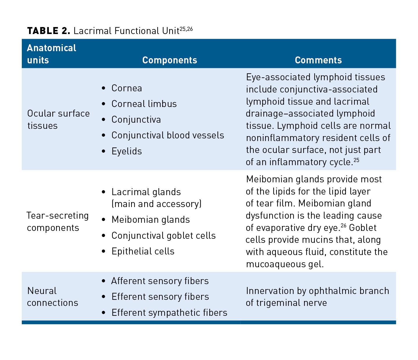
Normal physiology. Tear film is produced by the lacrimal functional unit (LFU), the anatomy of which includes ocular surface tissues, tear-secreting components, neural connections, and pathways for inflammatory and immune responses (Table 225,26). Stimulation of nerve endings in the cornea and nasal passages sends afferent impulses through the ophthalmic branch of the trigeminal nerve (cranial nerve V) to the central nervous system (brainstem and cerebral cortex). These sensory impulses regulate tear production and blinking. Efferent impulses arise in the superior salivatory nucleus and eventually reach the lacrimal gland via the lacrimal nerve.27
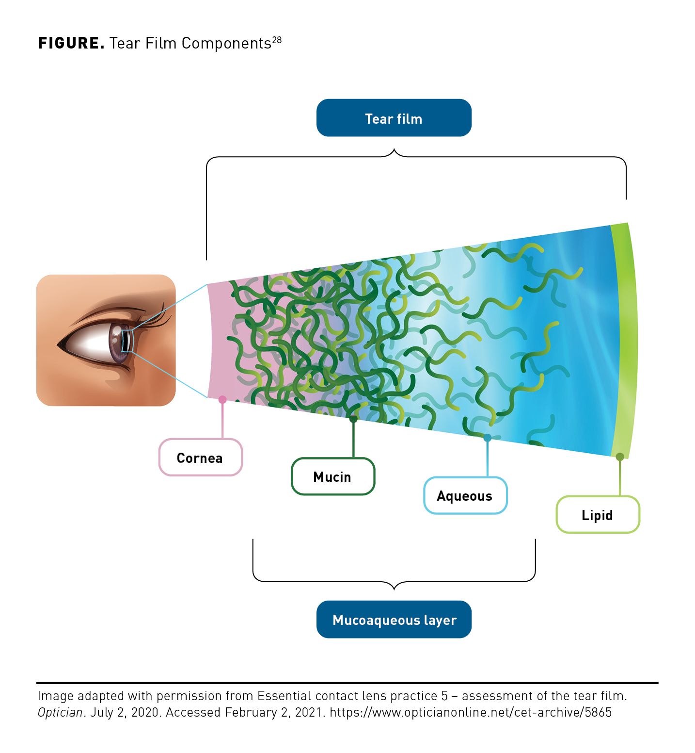
A healthy tear film lubricates the eyes for comfort, protects them from injury and infection, washes away foreign particles and debris from the turnover of epithelial cells, and maintains the smooth refractive surface necessary for clear vision. The traditional model of tear film incorporated 3 layers: an outer lipid layer covering an aqueous layer, both over a mucin layer covering epithelial cells.8,9 In recent years, the tear film model has been described as a complex blended 2-layer structure, comprised of a mucoaqueous layer and an outer lipid layer (Figure28). The entire tear film is only 2 to 6 µm thick.
Natural tears contain a biochemically complex mixture of electrolytes, at least 5 classes of lipids, more than 20 mucins, hundreds of peptides, and about 1500 proteins.9 Among the proteins are growth factors—including nerve growth factor, VEGF-A, and epidermal growth factor—that act as regulators of corneal growth and wound healing. Other proteins contained within natural tears have cytoprotective and immunologic functions.
Pathophysiology. The LFU is so tightly integrated that dysfunction in any single component can lead to dysfunction in others. There are numerous causes of LFU dysfunction, ranging from deficiencies in tear film components to blink abnormalities, but ocular surface homeostasis is lost when any LFU dysfunction occurs. The conceptual model of dry eye disease pathophysiology is a vicious circle, in which destabilization of the tear film results in tear hyperosmolarity; this is the hallmark of dry eye disease.27
Hyperosmolarity is a consequence of tear evaporation in both ADDE and EDE. However, although all forms of dry eye disease are evaporative, EDE is especially so and labeled hyperevaporative. In EDE, lacrimal function is normal, but evaporation is excessive; in ADDE, the rate of evaporation is normal, but lacrimal secretion is reduced. Either state results in hyperosmolarity. Environmental factors such as low humidity, high wind, and high temperatures can exacerbate tear film evaporation. Absent evaporation (or aqueous deficiency), hyperosmolarity does not occur.27
Hyperosmolarity triggers a cascade of signaling events in ocular surface epithelial cells, resulting in the release of inflammatory mediators (eg, interleukin 1, tumor necrosis factor-α) and proteases that damage epithelial cells and goblet cells of the ocular surface. Tear film instability then occurs, leading to tear film breakup that exacerbates hyperosmolarity, continuing the vicious circle that perpetuates the disease state.27
Although multiple published papers discuss dry eye disease, relatively few studies have examined its natural history and/or its clinical consequences.16 In general, if left untreated, dry eye disease may lead to corneal scarring or minor vision loss, although permanent loss of vision is relatively uncommon. Some cases resolve, but many patients require chronic therapy.
Diagnosis. An eye care or primary care provider will typically suspect a dry eye disease diagnosis on the basis of ocular symptoms reported by the patient (eg, complaints of blurred vision, red eyes, pain, burning, grittiness or irritation). Then, they will use various tests to confirm the diagnosis and identify the dry eye disease subtype to guide treatment. Disease symptoms and signs can vary over time and in response to numerous environmental factors such as systemic medications, wind exposure, cold weather, low humidity, and time of day. It should also be noted that in some cases, a patient with dry eye disease will present with symptoms but no signs, or with signs but no symptoms. If signs alone are present, treatment still may be warranted to prevent emergence of dry eye disease, especially if refractive surgery or contact lens use is planned, and significant signs in the absence of symptoms would suggest neurotropic keratitis. Symptoms in the absence of clinically observable signs suggest that neuropathic pain should be investigated.29
For eye care practitioners to use in clinical practice, DEWS II has developed triaging questions and several diagnostic tests. The recommended process begins with a differential diagnosis to exclude common conditions that mimic the signs and symptoms of dry eye disease. These include reactions to contact lens wear; adverse effects of systemic medications such as antihistamines, antidepressants, and anxiolytics; and others that include ocular infection, trauma, corneal dystrophies, and allergies. The diagnostic test battery includes screening with the 5-item Dry Eye Questionnaire or the 12-question Ocular Surface Disease Index© (© 1995 Allergan Inc. Irvine, CA. All rights reserved.), followed, if warranted, by a test for homeostasis markers, which includes either noninvasive tear breakup time or fluorescein tear breakup time, osmolarity testing, or ocular surface staining for assessing conjunctival, lid margin, and/or corneal damage.29-31
The diagnostic tests recommended by DEWS II for use in contemporary clinical practice are not necessarily the same as the tests used in clinical trials leading to FDA approval of a drug for treatment of dry eye disease (Table 332). For example, although the Schirmer test was once widely used to diagnose aqueous tear deficiency, DEWS II states that the variability and invasiveness of this test “precludes its use as a routine diagnostic test of tear volume, especially in cases with evaporative dry eye.”29
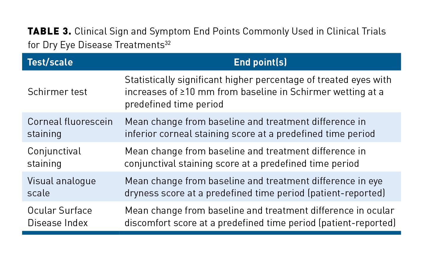
Nonetheless, until a new consensus on consistent and quantifiable diagnostic assays is reached, one of the main tests utilized to evaluate dry eye disease efficacy in clinical trials is the Schirmer test. The test involves placing a 5 mm × 35 mm strip of filter paper over the lower lid margin. Patients keep their eyes closed for 5 minutes, after which the length of the portion of the strip that has become wet is measured. Wetting of less than 5 mm of the strip commonly is regarded as the threshold for aqueous tear deficiency.33 The test can be performed with or without topical anesthesia.
Treatment. Because dry eye disease has been long regarded as a problem of tear insufficiency, tear replacement therapy with ocular lubricants (“artificial tears”) has become the mainstay of therapy. Although treatment algorithms exist, as described below, currently available therapeutics treat the symptoms rather than the underlying cause of the disease.
Algorithm. In its report for the DEWS II Workshop, the Management and Therapy Committee published a 4-step algorithm for management of dry eye disease once its subtype had been determined.15 If patients fail to respond to a step or if their symptoms are more severe, advancement to the next step is recommended, retaining any previous therapy as warranted.
- Step 1: Initial measures involve educating the patient about dry eye disease and its management, treatment, and prognosis; addressing any local environmental factors; identifying and eliminating or modifying any systemic or topical medications that may contribute to dry eye disease symptoms; recommending ocular lubricants; and recommending lid hygiene and warm compresses.
- Step 2: Additional options include nonpreserved ocular lubricants, tear conservation with punctal occlusion or moisture chamber goggles, in-office heating and expression of meibomian glands or in-office pulsed light therapy for meibomian gland dysfunction (in equipped ophthalmologists’ offices), and the following prescription drugs: topical antibiotic or antibiotic/steroid combinations to treat blepharitis, if present; topical limited-duration corticosteroids (longer-duration corticosteroids are reserved for step 4); topical nonglucocorticoid immunomodulatory drugs (eg, cyclosporine); topical LFA-1 antagonists (eg, lifitegrast); and oral macrolide or tetracycline antibiotics. (Another component of step 2 is topical secretagogues; however, they are not available in the United States.)
- Step 3: Additional options include autologous/allogenic serum eyedrops and therapeutic contact lenses. (Another component of step 3 is oral secretagogues; however, they are not available in the United States.)
- Step 4: Additional options include longer-duration topical corticosteroids, amniotic membrane grafts, and surgical punctal occlusion, among other surgical approaches.
Unmet Needs
Despite the explosion of knowledge about dry eye disease during the last 2 decades, much remains to be learned about dry eye disease in all areas—epidemiology, biochemistry and physiology, diagnosis, and treatment, among others.
In epidemiology, more studies are needed about dry eye disease in populations younger than 40 years, the natural history of dry eye disease with and without treatment, risk factors, indirect costs stemming from lost productivity, and cost savings stemming from treatment.18
Identification of biomarkers for dry eye disease could help clinicians to screen for and diagnose dry eye disease, as well as monitor response to treatment, and some biomarkers could become targets of treatment or treatments in themselves.1 Willcox and colleagues (2017) envision the development of a precision medicine approach to dry eye disease in which identification of a deficiency in 1 or more functionally important proteins in tear film guides topical treatment to address the deficiency.9
At the level of ocular innervation, little is known about ocular pain. It would be helpful to differentiate the nociceptive pain in dry eye disease from neuropathic ocular pain, and more information is needed about management of neuropathic pain detected during investigation of dry eye disease.34 Much more knowledge also is needed about neural regulation of meibomian gland secretion and mucin release. Increased knowledge of these topics could facilitate development of new treatments and help clinicians to monitor patients’ response to therapy.1
In the treatment of dry eye disease, evidence about the relative value of currently available treatment options is lacking. Given that over-the-counter (OTC) ocular lubricants are used so often in the early treatment of dry eye disease and that a great many of these products are on the market, randomized controlled trials comparing the efficacy of products are especially needed, including studies of products with and without lipids in treating EDE and ADDE; the effects of OTC lubricants on tear film osmolarity; and the length of time OTC lubricants must be used to effect beneficial changes.12
Panel Discussion
Payers and clinicians discussed how their knowledge of dry eye disease has increased over the years, resulting in changes in their attitudes toward its management and treatment. Clinicians said they sometimes encountered barriers to treatment during interactions with managed care organizations (MCOs), but payers generally did not view the class of drugs for dry eye disease as one that warranted significant management. Payers and clinicians shared a common view that their peers would benefit from additional education to appreciate the existing unmet need for therapeutic options that may address the underlying causes of dry eye disease.
Evolving Attitudes Toward Disease State and Treatment
Clinician perspective: Although some specialty clinicians initially regarded dry eye disease as a nuisance, they have come to appreciate dry eye disease as a true disease that substantially degrades QOL and that, in its more severe forms, can be very debilitating. It is among the most common reasons patients visit eye care practitioners. Clinical consequences may include blurred vision, red eyes, chronic pain, burning, and irritation. Dry eye disease may lead to permanent long-term damage to meibomian glands and the ocular surface. It also is responsible for many cases of refractive inaccuracies after cataract surgery, and it is a leading reason for contact lens dropout. Clinician panelists estimated that at least 25 to 30 million people in the United States have some form of clinically significant dry eye. One clinician mentioned study results finding that patients with severe dry eye disease rank its overall impact as similar to that of the discomfort and distress experienced by patients who have severe angina or are undergoing kidney dialysis.35
For years, even as clinicians, we originally thought of dry eye disease as a
nuisance, kind of like chapped lips.”
— Optometrist, dry eye disease clinic
In the last decade, especially, clinicians have come to appreciate dry eye disease as a complex condition, multifactorial in its diagnosis and treatment. Clinicians also shared that the most common limitation of currently available treatments is their slow onset of action, requiring weeks, if not months, of use before patients perceive a benefit. Because dry eye disease is now known as a disease where symptoms and signs do not correlate to the severity of the disease, they said it has been difficult for researchers to create good clinical studies that yield positive results, which has consequently limited research and development of new therapeutics.
They noted that even patients may perceive dry eye disease as not being a “big deal”—until it becomes one, mostly by greatly impeding their QOL. When dry eye disease makes every activity difficult, it becomes far more than a nuisance.
The clinicians said that if 16 million patients have received a diagnosis of dry eye disease, the diagnosis may have been missed in a comparable number of adults in the United States because eye care practitioners have not always thought to suspect dry eye disease. Further, because signs and symptoms of dry eye disease don’t necessarily correlate, a clinician may fail to ask the patient about symptoms even though signs are evident.
… the bigger factors are effects on vision—not to the point of blindness, but
really affecting [patients’] functioning.”
— Ophthalmologist
Optometrist and ophthalmologist panelists both mentioned that because about 10% of dry eye disease is purely aqueous, about 45% purely evaporative, and about 40% mixed, treatment of a large percentage of patients with dry eye disease involves more than addressing just the aqueous component. As such, they said, artificial tears might lower osmolarity, and although the products could have some mucin-like components, along with some electrolytes and pH balance, artificial tears have none of the proteins or mucins found in natural tears.
Payer perspective: Like the clinicians, the managed care representatives had the view that dry eye disease has been perceived as a nuisance or an irritant instead of as a clinical disease, and many times the category of dry eye disease treatments is rushed through pharmacy and therapeutics committee discussions. One payer said that although different levels of dry eye disease severity are recognized, treatments for dry eye disease definitely are not on his organization’s radar screen because dry eye disease is a relatively low-cost condition.
Many times, we rush this category in the P&T [pharmacy and therapeutics
committee] discussion [as it has] a big lifestyle component. Understanding the etiology and perhaps the unmet need [may lead] to more robust discussions.”
— Managed care organization executive
During the panel discussion and upon hearing the clinicians’ perspectives, payers commented that dry eye disease may be more complex than they previously thought. They said it is important for payers to understand that treatment involves more than just replacing lost water. Because lipids and proteins in natural tear film obviously perform various functions, it’s “sophomoric” to think we can replace all the lost function with eyedrops, it was said.
In addition, payers said it is important to know that dry eye disease has many components, one of which is inflammation, and that there is an unmet need for targeting the underlying reasons for the lack of tear production.
Approaches to Coverage of Dry Eye Disease Treatments
Clinicians strive to prescribe a treatment regimen tailored to meet a patient’s specific medical needs but may adjust the treatment plan if clinical considerations or affordability present a barrier to adherence. Most payers don’t regard prescription drugs for dry eye disease as a high priority for management, but some MCOs do use step edits and prior authorization in efforts to ensure appropriate use and manage drug spend.
Clinician perspective: The clinicians identified prior authorization requests, variations in patients’ insurance coverage, and patients’ out-of-pocket expenses as the chief barriers encountered when they prescribe prescription drugs for dry eye disease.
Ideally, said the clinicians, they try to use a multifactorial approach to identify the best course of treatment for their patients, according to the type of disease and risk factors. Using their knowledge of the drugs that insurers in their region cover, the clinicians often initiate therapy with whatever drug the insurer will allow and what the patient can afford. If the clinical need is to control inflammation, and one anti-inflammatory drug will cost the patient $300 a month but another anti-inflammatory costs $50, the decision is easy. In the absence of knowledge of insurance coverage, clinicians said they may prescribe the product they believe may be most appropriate and provide the patient with 1 or more options to initiate treatment. After the patient’s experience with the initial treatment, a provider would explore other options if the outcome, tolerability, or affordability becomes a concern.
Clinicians encounter prior authorization requests frequently, but only a small percentage of these result in denials. Sometimes, even if a drug is approved, patients can’t afford the co-pay. It doesn’t matter to the patient that the co-pay is only a small percentage of the true cost of the product for the monthly supply.
Payer perspective: The managed care representatives reported different approaches by their organizations to cover prescription drugs for dry eye disease. Most said dry eye disease drugs were not high-priority products, given their relatively low cost, especially in comparison with the much more expensive targeted products used to treat life-threatening diseases such as cancer or inflammatory conditions.
This isn’t a category that rises to significant management activity.”
— Managed care organization executive
One MCO executive reported having dry eye disease products on formulary but no prior authorization or step edits on them. Several payer participants suggested that dry eye disease is not something their organizations are trying to manage at the moment; however, their approach to dry eye disease may change as new dry eye disease products come to market. Other payer representatives stated that their organizations use step edits and prior authorization to manage dry eye disease prescription drugs, in efforts to ensure appropriate utilization and managed drug spend. Although not in the category of specialty pharmaceuticals, currently prescribed dry eye disease prescription drugs still are sufficiently expensive to warrant some management by some MCOs.
Unmet Needs in Dry Eye Disease
In the eyes of the clinicians, the relatively long time—weeks or months—required for many patients to experience improvement is a challenge with some prescribed therapeutics for dry eye disease. A potential shortcoming among many of their peers across the country, the 2 clinician panelists said, is a focus on treatment of inflammation alone, to the exclusion of the factor(s) responsible for the inflammation. Eye care practitioners would benefit from further education about dry eye disease’s multifactorial nature.
Clinician perspective: A challenge they have experienced with their patients taking some of the currently available prescription drugs for dry eye disease, the clinicians said, is the length of time it takes for benefits to become apparent.
Clinicians pointed to an insufficient understanding among some eye care and primary care providers of how to manage dry eye disease. They said an unmet need involves educating clinicians to address all components of dry eye disease, not just inflammation, and providing information to clinicians that will facilitate a change in their current treatment approaches. Some patients who have discontinued dry eye disease treatment may have done so because they are discouraged and think they have tried everything and have no other options.
The panelists explained that in EDE, for example, if the lipid layer is deficient, the patient typically has obstructed glands and biofilm or blepharitis. This patient has inflammation and a tear film issue because of lack of the right mucins, proteins, and lipids. When such patients are referred to a specialist in dry eye disease, they are suffering.
Another unmet need, the panelists said, is greater understanding among eye care and primary care providers of the qualitative and quantitative nature of EDE, which is a relatively new field. In the absence of a prescription therapy specifically for EDE, there hasn’t been much education of providers. This is a problem, participating clinicians felt, because even moderate-to-severe EDE is rampant and getting worse.
An obvious need, the clinicians said, is for therapies that could address the underlying issues of dry eye disease beyond inflammation. Clinicians’ desire for alternatives does not imply negation of anti-inflammatories. There are times when, if inflammation, obstructed meibomian glands, and tear film are controlled, patients can stay on anti-inflammatory medications longer because those drugs are helping. But treating just the result of obstructed glands or the result of biofilm or the result of inadequate tear film—inflammation—is not sufficient for many patients and may exacerbate the condition in the long term. If the cause of inflammation is not addressed, more meibomian glands can be lost, drop out, or atrophy, and more damage and scarring can ensue. Clinicians suggested that a mechanism to address the root cause of dry eye disease and restore the ability of components of the LFU to produce normal tear film would be optimal. Currently, eye care practitioners do this more crudely than they would like, with compresses, lid debridement, and anti-inflammatories that improve tear film as best they can. Eventually, if medications and therapeutics that target each of these components are developed, patient outcomes may improve. Substantial progress can’t necessarily be made with 1 drug that addresses 1 component.
Clinician panelists also felt that better ways to qualify and quantify QOL measures of dry eye disease are necessary. The QOL perspective is important to patients and health care providers, and panelists hope to elevate payers’ understanding of the effect of dry eye disease on patients’ lives. Clinicians regularly see how dry eye disease adversely affects patients, but that knowledge is not translated to payers because there is a lack of published evidence.
Payer perspective: Over the last decade, the payers said, a better understanding of dry eye disease has been acquired, with respect to its different etiologies and manifestations and the consequences of not treating dry eye disease appropriately or at all. Payers acknowledged that the understanding of dry eye disease is evolving but that much more information and education on the disease are needed for perceptions to change.
The covered drugs in this category may not be working for everyone,
as evidenced by the numbers of people who have tried treatment only
to discontinue it. What are the downstream issues and effects? Those
are key points to understand.”
— Managed care organization executive
Payers said a need for different therapy options appears to still exist because many patients do not respond to current therapies. One payer provided a summary of their MCO’s actual data regarding reasons for members’ discontinuation of prescription drugs for dry eye disease (Table 4). “Just looking at the percentages without clinical improvement tells me that we need something different,” the payer said. Another payer expressed a hope to see more studies become available to their peers in MCOs about the impact of dry eye disease on QOL and about clinical measures to treat it.
In addition to alternative therapy options that would include but not be limited to drug treatments, payers said they would like a management algorithm that could help them understand the appropriate situations for various options.

Conclusions
Although some eye care practitioners and payers may have historically regarded dry eye disease as more of a nuisance than an actual disease or condition, current thinking is evolving to recognize dry eye disease as a common, complicated, multifactorial, and underdiagnosed disease that impairs the lives of millions of adults in the United States. Dry eye disease can greatly affect patients’ ocular and overall health, as well as impact their QOL, and more studies to quantify these impacts are needed. Payers and clinicians alike see a need for additional therapeutic options that could address the underlying causes of dry eye disease and have the potential to positively impact patient outcomes. Education about disease etiology, underlying causes, and therapeutics’ mechanisms of action is essential for health care decision makers and providers to change their perceptions and approaches to formulary management and treatment, respectively.
Acknowledgments
The authors gratefully acknowledge Jack McCain for editorial assistance and would like to thank their colleagues on the Evolving Knowledge of the Unmet Needs in Dry Eye Disease virtual roundtable panel for their support.
Author affiliations: Magellan Rx (formerly), Salt Lake City, UT (JDD); Kentucky Eye Institute, Lexington, KY, and Kentucky College of Optometry, Pikeville, KY (PMK); National health plan/pharmacy benefit manager (MEM); No affiliation/independent managed care pharmacy consultant, San Marcos, CA (DR).
Funding source:The Evolving Knowledge of the Unmet Needs in Dry Eye Disease virtual roundtable and production of this report were sponsored by Oyster Point Pharma.
Author disclosures: Dr Karpecki has served as a consultant or paid advisory board member for AbbVie Inc; Bausch + Lomb Inc; Kala Pharmaceuticals; Novartis Inc; Oyster Point Pharma, Inc; and Sun Pharmaceutical Industries Ltd; he also reports receipt of lecture fees from Bausch + Lomb Inc, Dompé, Eyevance Pharmaceuticals LLC, Kala Pharmaceuticals, Mallinckrodt Pharmaceuticals, Neurolens, Osmotica Pharmaceutical, and Sun Pharmaceutical Industries Ltd. Dr Reissman has served as a consultant or paid advisory board member and received honoraria for participation in blinded market research interviews on dry eye disease. Drs Dunn and Meske report no relationship or financial interest with any entity that would pose a conflict of interest with the subject matter of this supplement.
Authorship information: Concept and design (PMK); analysis and interpretation of data (JDD, PMK, MEM, DR); drafting of the manuscript (JDD, MEM, DR); critical revision of the manuscript for important intellectual content (JDD, PMK, MEM, DR); and administrative, technical, or logistic support (MEM).
Address correspondence to: Paul M. Karpecki, OD, Kentucky Eye Institute, 601 Perimeter Dr, Ste 100, Lexington, KY 40517. Email: paul@karpecki.com
References
1. Clayton JA. Dry eye. N Engl J Med. 2018;378(23):2212-2223. doi:10.1056/NEJMra1407936
2. Wang MTM, Muntz A, Lim J, et al. Ageing and the natural history of dry eye disease: a prospective registry-based cross-sectional study. Ocul Surf. 2020;18(4):736-741. doi:10.1016/j.jtos.2020.07.003
3. Miljanovic´ B, Dana R, Sullivan DA, Schaumberg DA. Impact of dry eye syndrome on vision-related quality of life. Am J Ophthalmol. 2007;143(3):409-415. doi:10.1016/j.ajo.2006.11.060
4. McDonald M, Patel DA, Keith MS, Snedecor SJ. Economic and humanistic burden of dry eye disease in Europe, North America, and Asia: a systematic literature review. Ocul Surf. 2016;14(2):144-167. doi:10.1016/j.jtos.2015.11.002
5. Lemp MA. Report of the National Eye Institute/Industry Workshop on clinical trials in dry eyes. CLAO J. 1995;21(4):221-232.
6. The definition and classification of dry eye disease: report of the Definition and Classification Subcommittee of the International Dry Eye Workshop (2007). Ocul Surf. 2007;5(2):75-92. doi:10.1016/s1542-0124(12)70081-2
7. Craig JP, Nelson JD, Azar DT, et al. TFOS DEWS II Report Executive Summary. Ocul Surf. 2017;15(4):802-812. doi:10.1016/j.jtos.2017.08.003
8. Pflugfelder SC, Stern ME. Biological functions of tear film. Exp Eye Res. 2020;197:108115. doi:10.1016/j.exer.2020.108115
9. Willcox MDP, Argüeso P, Georgiev GA, et al. TFOS DEWS II tear film report. Ocul Surf. 2017;15(3):366-403. doi:10.1016/j.jtos.2017.03.006
10. Thoft RA, Friend J. Biochemical transformation of regenerating ocular surface epithelium. Invest Ophthalmol Vis Sci. 1977;16(1):14-20.
11. Stern ME, Beuerman RW, Fox RI, Gao J, Mircheff AK, Pflugfelder SC. The pathology of dry eye: the interaction between the ocular surface and lacrimal glands. Cornea. 1998;17(6):584-589. doi:10.1097/00003226-199811000-00002
12. Jones L, Downie LE, Korb D, et al. TFOS DEWS II management and therapy report. Ocul Surf. 2017;15(3):575-628. doi:10.1016/j.jtos.2017.05.006
13. Behrens A, Doyle JJ, Stern L, et al; Dysfunctional Tear Syndrome Study Group. Dysfunctional tear syndrome: a Delphi approach to treatment recommendations. Cornea. 2006;25(8):900-907. doi:10.1097/01.ico.0000214802.40313.fa
14. Villani E, Marelli L, Dellavalle A, Serafino M, Nucci P. Latest evidences on meibomian gland dysfunction diagnosis and management. Ocul Surf. 2020;18(4):871-892. doi:10.1016/j.jtos.2020.09.001
15. Jones L, Downie LE, Korb D, et al. TFOS DEWS II management and therapy report. Ocul Surf. 2017;15(3):575-628. doi:10.1016/j.jtos.2017.05.006.
16. Nelson JD, Craig JP, Akpek EK, et al. TFOS DEWS II introduction. Ocul Surf. 2017;15(3):269-275. doi:10.1016/j.jtos.2017.05.005
17. Craig JP, Nichols KK, Akpek EK, et al. TFOS DEWS II Definition and Classification Report. Ocul Surf. 2017;15(3):276-283. doi:10.1016/j.jtos.2017.05.008
18. Stapleton F, Alves M, Bunya VY, et al. TFOS DEWS II epidemiology report. Ocul Surf. 2017;15(3):334-365. doi:10.1016/j.jtos.2017.05.003
19. Paulsen AJ, Cruickshanks KJ, Fischer ME, et al. Dry eye in the Beaver Dam Offspring Study: prevalence, risk factors, and health-related quality of life. Am J Ophthalmol. 2014;157(4):799-806. doi:10.1016/j.ajo.2013.12.023
20. Farrand KF, Fridman M, Stillman IÖ, Schaumberg DA. Prevalence of diagnosed dry eye disease in the United States among adults aged 18 years and older. Am J Ophthalmol. 2017;182:90-98. doi:10.1016/j.ajo.2017.06.033
21. Wang MTM, Muntz A, Lim J, et al. Ageing and the natural history of dry eye disease: a prospective registry-based cross-sectional study. Ocul Surf. 2020;18(4):736-741. doi:10.1016/j.jtos.2020.07.003
22. Akkaya S, Atakan T, Acikalin B, Aksoy S, Ozkurt Y. Effects of long-term computer use on eye dryness. North Clin Istanb. 2018;5(4):319-322. doi:10.14744/nci.2017.54036
23. Choi JH, Li Y, Kim SH, et al. The influences of smartphone use on the status of the tear film and ocular surface. PLoS One. 2018;13(10):e0206541. doi:10.1371/journal.pone.0206541
24. Jaiswal S, Asper L, Long J, Lee A, Harrison K, Golebiowski B. Ocular and visual discomfort associated with smartphones, tablets and computers: what we do and do not know. Clin Exp Optom. 2019;102(5):463-477. doi:10.1111/cxo.12851
25. Knop E, Knop N. Influence of the eye-associated lymphoid tissue (EALT) on inflammatory ocular surface disease. Ocul Surf. 2005;3(suppl 4):S180-S186. doi:10.1016/s1542-0124(12)70251-3
26. Bron AJ, Tiffany JM. The contribution of meibomian disease to dry eye. Ocul Surf. 2004;2(2):149-165. doi:10.1016/s1542-0124(12)70150-7
27. Bron AJ, de Paiva CS, Chauhan SK, et al. TFOS DEWS II pathophysiology report. Ocul Surf. 2017;15(3):438-510. doi:10.1016/j.jtos.2017.05.011. Published correction appears in Ocul Surf. 2019;17(4):842.
28. Essential contact lens practice 5 – assessment of the tear film. Optician. July 2, 2020. Accessed February 2, 2021. https://www.opticianonline.net/cet-archive/5865
29. Wolffsohn JS, Arita R, Chalmers R, et al. TFOS DEWS II diagnostic methodology report. Ocul Surf. 2017;15(3):539-574. doi:10.1016/j.jtos.2017.05.001
30. Schiffman RM, Christianson MD, Jacobsen G, Hirsch JD, Reis BL. Reliability and validity of the Ocular Surface Disease Index. Arch Ophthalmol. 2000;118(5):615-621. doi:10.1001/archopht.118.5.615
31. Geerling G, Tauber J, Baudouin C, et al. The International Workshop on Meibomian Gland Dysfunction: report of the Subcommittee on Management and Treatment of Meibomian Gland Dysfunction. Invest Ophthalmol Vis Sci. 2011;52(4):2050-2064. doi:10.1167/iovs.10-6997g
32. Nichols KK, Evans DG, Karpecki PM. A comprehensive review of the clinical trials conducted for dry eye disease and the impact of the vehicle comparators in these trials. Curr Eye Res. Published online November 25, 2020. doi:10.1080/02713683.2020.1836226
33. Kloosterboer A, Dermer HI, Galor A. Diagnostic tests in dry eye. Expert Rev Ophthalmol. 2019;14(4-5):237-246. doi:10.1080/17469899.2019.1657833
34. Belmonte C, Nichols JJ, Cox SM, et al. TFOS DEWS II Pain and Sensation Report. Ocul Surf. 2017;15(3):404-437. doi:10.1016/j.jtos.2017.05.002
35. Buchholz P, Steeds CS, Stern LS, et al. Utility assessment to measure the impact of dry eye disease. Ocul Surf. 2006;4(3):155-161. doi:10.1016/s1542-0124(12)70043-5

