- Center on Health Equity & Access
- Clinical
- Health Care Cost
- Health Care Delivery
- Insurance
- Policy
- Technology
- Value-Based Care
Diaphragm Ultrasound Imaging Showcases Promise for Respiratory Assessments in SMA
In this prospective study, respiratory monitoring of patients with spinal muscular atrophy (SMA) benefited from the use of diaphragm ultrasound imaging.
Diaphragm ultrasound imaging (DUI) proved to be a valuable tool for disease and treatment assessment in patients with spinal muscular atrophy (SMA), according to a study published in Muscle & Nerve.
In SMA, characteristic muscle atrophy and weakness commonly affect ventilatory muscles as well. Respiratory impairment in these patients varies in severity but can necessitate ventilation. Previous research has found, however, that the diaphragm is seemingly less affected by the survival motor neuron 1 (SMN1) gene malfunction.
The diaphragm’s resistance to the SMN1 mutation is believed to play a part in the preserved respiratory function observed in some SMA types that result in drastically affected motor function.
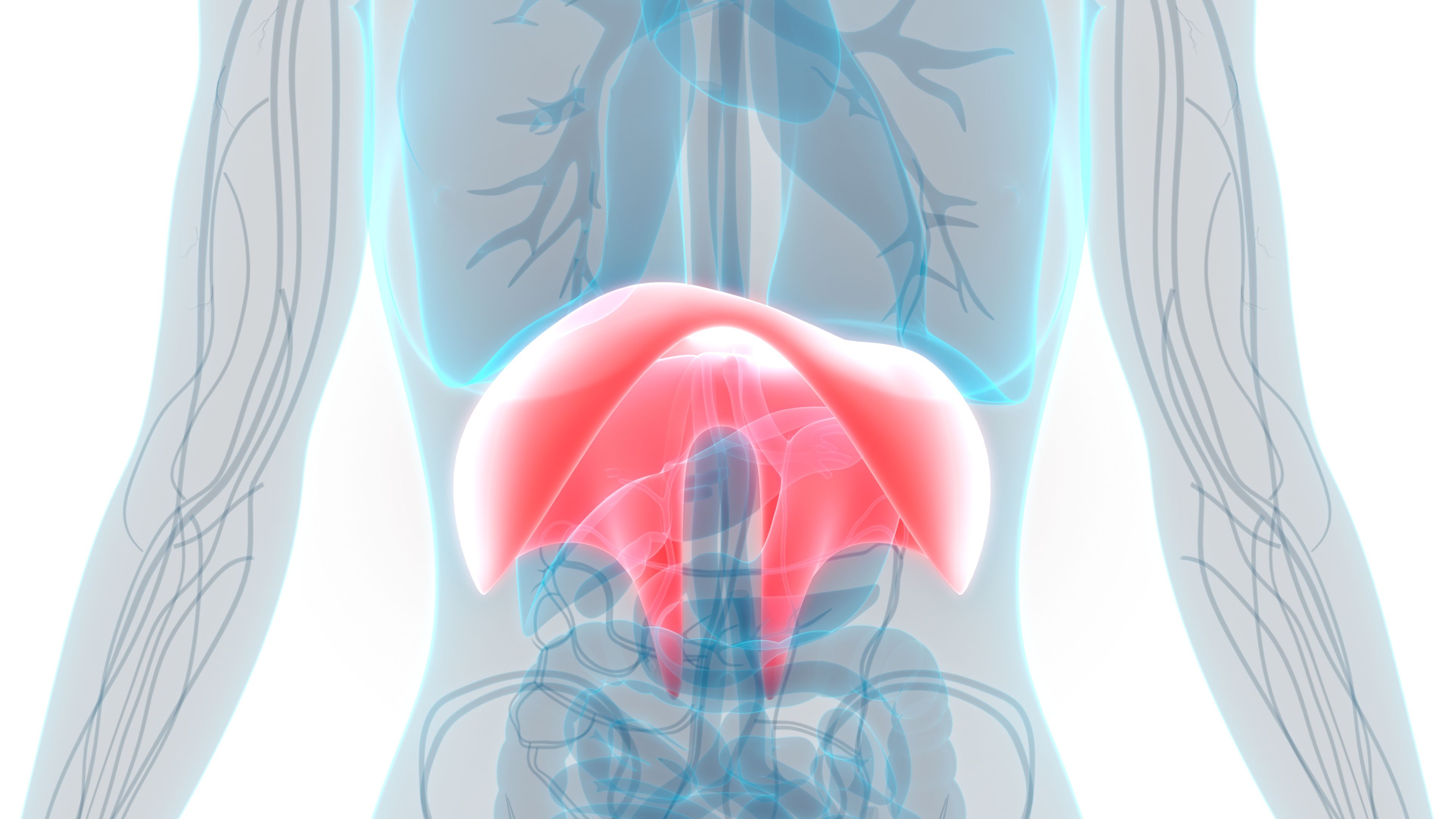
Human Diaphragm | Image credit: magicmine - stock.adobe.com

The authors note that, since cases of ventilatory damage are often fatal, autopsy studies and postmortem tissues have provided a large amount of the known pathoanatomic information. Because there are not sufficient data on respiratory outcomes in adult patients with SMA, researchers conducted a study using DUI to evaluate diaphragm functioning and responses to nusinersen treatment in this population.
Between September 2017 and September 2021, data were gathered from the Department of Neurology of the University Hospital Carl Gustav Carus Dresden, Germany, to identify adult patients with SMA. A diaphragm thickening fraction (DFT) was utilized and any results less than 20% were considered reduced. DFT values were referenced against study results from Boussuges et al, which demonstrated a mean right-diaphragm thickness of 1.9 mm for women and 2.1 mm for men, and a left-side mean of 1.7 and 2.0 mm, respectively. Diaphragm excursion was also measured and healthy individuals were considered to range from 17 to 20 mm. The authors noted that some measurements were missing due to constraints in the treatment period or changes in treatment.
Twenty-four adults with SMA were identified and had DUIs repeated after 15.5, 26.6, and 38.3 months on average. Preceding nusinersen treatment, DTF, and excursion decreases were seen in 25% of participants and diaphragm thickness increased for many. In these results, the authors found that diaphragm thickness was not indicative of atrophy in patients; however, these structural changes did suggest a degree of diaphragmatic dysfunction. Furthermore, they witnessed dramatic differences in spirometric parameters between those with SMA types 2 and 3. These differences were not seen in DUI results, which suggests that ventilatory impairment was associated with respiratory muscles rather than the diaphragm.
During nusinersen treatment, patients experienced improved thickening and excursion outcomes. Spirometric parameters were stable throughout and all but 1 patient were able to return to normal diaphragmatic motility.
These findings demonstrate the importance of respiratory monitoring in patients with SMA. Identifying diaphragmatic changes over the course of SMA is essential for anticipating respiratory failure in these patients. In this study, DUI proved to be a complimentary tool in this investigation and the authors believe it should be considered to help guide the management of patients with SMA to provide the best care possible. Because of their limited sample size, the authors concluded by emphasizing the need to validate these findings in trials with larger cohorts, multiple centers, and longer study periods.
Reference
Freigang M, Langner S, Hermann A, Günther R. Impaired diaphragmatic motility in treatment-naïve adult patients with spinal muscular atrophy improved during nusinersen treatment. Muscle Nerve. 2023;68(3):278-285. doi:10.1002/mus.27938
